- Anatomical terminology
- Skeletal system
- Joints
- Classification of joints
- Joints of skull
- Joints of spine
- Joints of lower limb
- Muscles
- Heart
- Blood vessels
- Lymphatic system
- Nervous system
- Respiratory system
- Digestive system
- Urinary system
- Female reproductive system
- Male reproductive system
- Endocrine glands
- Eye
- Ear
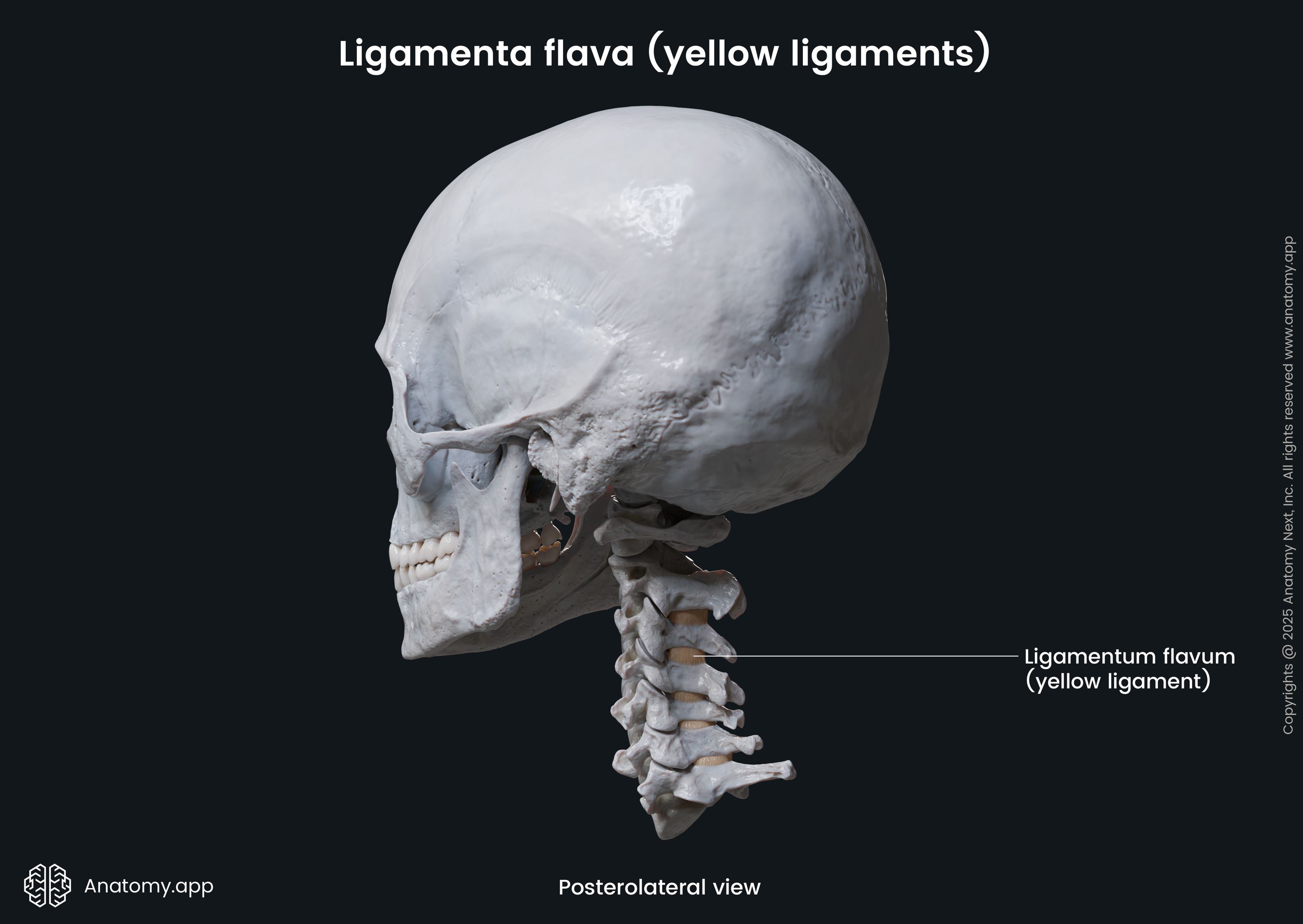
Ligamenta flava
The ligamenta flava (singular in Latin: ligamentum flavum) are highly elastic and broad paired fibrous bands that extend between laminae of adjacent vertebrae. They are thinner in the cervical region and become progressively thicker towards the lumbar spine. These ligaments are also known as the yellow ligaments as Ligamenta flava in Latin means “yellow ligaments”. This is because they contain high amounts of elastic tissue that has a yellow color, and therefore, these ligaments actually appear yellowish.
Ligamenta flava anatomy
The ligamenta flava are located throughout most of the spine. They are the thinnest in the cervical spine and gradually become thicker towards the lumbar spine. Similarly, as the distance between the laminae towards the thoracic region increases, the ligamenta flava also increase in length. The ligaments extend from the first two cervical vertebrae (C1 / C2) superiorly and terminate at the sacral region inferiorly - at the level of the fifth lumbar and first sacral vertebrae (L5 / S1).
The posterior atlanto-occipital membrane is an analog of the ligamenta flava at the level of the atlanto-occipital (C1 - occiput) junction. At the atlanto-axial joint level (C1 - C2), the ligamenta flava typically are thin and membranous. Moreover, some authors state that the ligamenta flava at the C1 - C2 level is rather the posterior atlanto-axial membrane. Here the ligamentum flavum is usually pierced by the second cervical (C2) spinal nerve.
Each intervertebral segment contains two ligamenta flava - one right and one left. Both connect laminae of two adjacent vertebrae. The fibers of each ligamentum flavum extend from the anterior surface and inferior margin of the upper lamina to the posterior surface and superior margin of the lower lamina.
The ligamenta flava extend laterally to the facet (zygapophyseal) joints and fuse together with their fibrous capsules. Moreover, each ligamentum flavum forms the anterior aspect of the facet joint capsule. Here it borders with the intervertebral foramina and its neurovascular components. The laminae of the vertebral arches and the ligamenta flava form the posterior wall of the spinal canal.
In the midline of the vertebral arch, the right and left ligamenta flava fuse together, as well as in the posterior direction, they merge with the interspinous ligament. Nevertheless, small gaps are present between merged ligamenta flava. These gaps transmit veins that unite the posterior internal (epidural) vertebral venous plexus with the posterior external vertebral venous plexus.
Ligamenta flava composition
The ligamenta flava consist predominantly of elastic fibers (around 80%). However, they also contain collagen fibers (around 20%). These ligaments show one of the highest amounts of elastic tissue in the human body. This unique feature allows the yellow ligaments to constrict in order for the spine to return to its anatomical position. The main component of the elastic tissue is called elastin, which contributes to the characteristic yellowish color of the ligaments.
Ligamenta flava functions
The ligamenta flava limit flexion of the spine and resist separation of the laminae during flexion. They aid in extension of the spine and return it to its anatomical position after flexion. Therefore, the ligamenta flava contributes to maintaining an upright body posture and help to preserve the curvatures of the spine. They also provide a smoother posterior lining of the vertebral canal, thus preventing contact between bony and adjacent neurovascular structures during extension.
Ligamenta flava issues
The ligamenta flava are very important structures responsible for the preservation of the integrity of the spine. These ligaments may become hypertrophied (abnormally enlarged) with aging or after traumas. They usually lose their elasticity, increase in thickness, calcify or get infiltrated with fat tissue. All these degenerative changes typically correspond to hypertrophy and arthritis of the zygapophyseal (or facet) joints. And finally, these processes can further result in the narrowing of the spinal canal and compression of its contents.
References:
- Adam, A., Dixon, A., & Gillard, J. (2020). Grainger & Allison’s Diagnostic Radiology: 2-Volume Set (7th ed.). Elsevier.
- Cramer, D. C., & Gregory, D. (2014). Clinical Anatomy of the Spine, Spinal Cord, and ANS, 3e 3rd (third) Edition by Cramer DC PhD, Gregory D., Darby PhD, Susan A. published by Mosby (2014) (3rd ed.). Mosby.
- Gray, H., & Carter, H. (2021). Gray’s Anatomy (Leatherbound Classics) (Leatherbound Classic Collection) by F.R.S. Henry Gray (2011) Leather Bound (2010th Edition). Barnes & Noble.
Anatomy.app
Contact information
- For questions regarding business inquiries. Please contact:
- info@anatomy.app