- Anatomical terminology
- Skeletal system
- Joints
- Classification of joints
- Joints of skull
- Joints of spine
- Joints of lower limb
- Muscles
- Heart
- Blood vessels
- Lymphatic system
- Nervous system
- Respiratory system
- Digestive system
- Urinary system
- Female reproductive system
- Male reproductive system
- Endocrine glands
- Eye
- Ear
Posterior longitudinal ligament
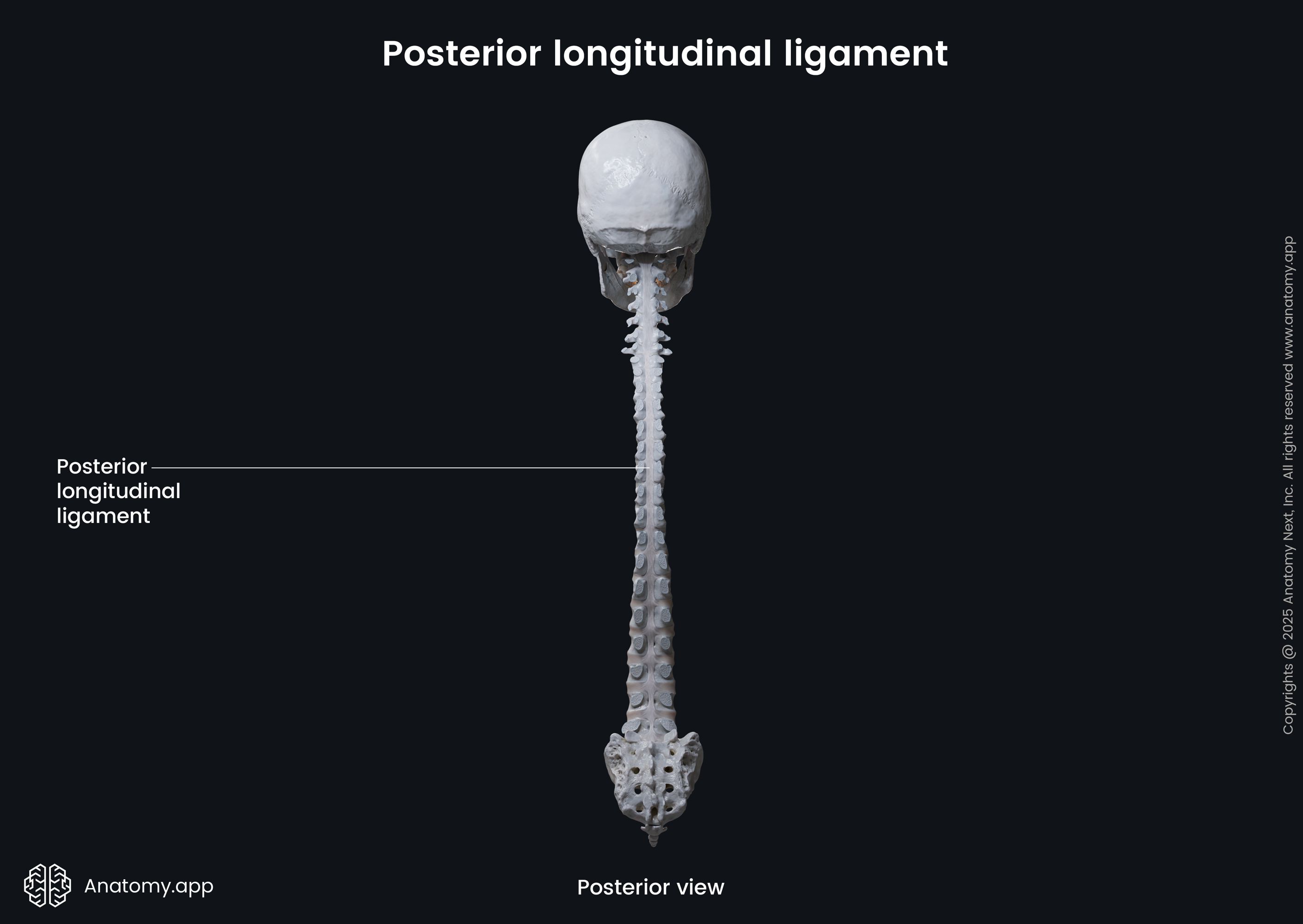
The posterior longitudinal ligament (Latin: ligamentum longitudinale posterius) is a strong band that runs posteriorly along the entire length of the vertebral column. Together with other primary ligaments, it contributes to the stability of the spinal structures.
Posterior longitudinal ligament anatomy
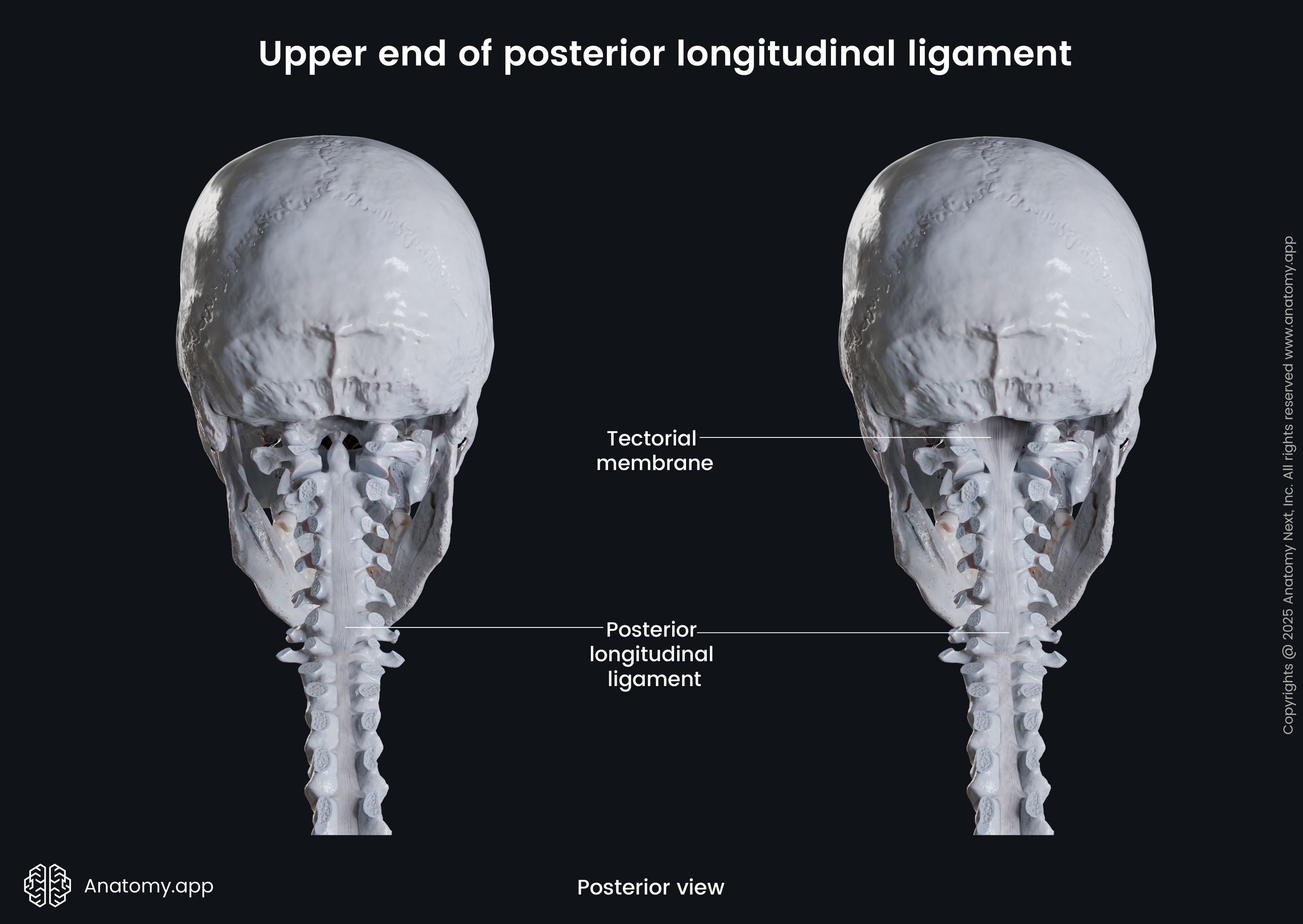
The posterior longitudinal ligament attaches to the posterior surfaces of the vertebral bodies and their respective intervertebral discs. Essentially, it is located within the vertebral canal on its anterior wall. The posterior longitudinal ligament runs from the body of the axis (C2) down to the sacral vertebrae (sacrum). This ligament is an inferior continuation of the tectorial membrane, which connects the axis to the base of the skull.
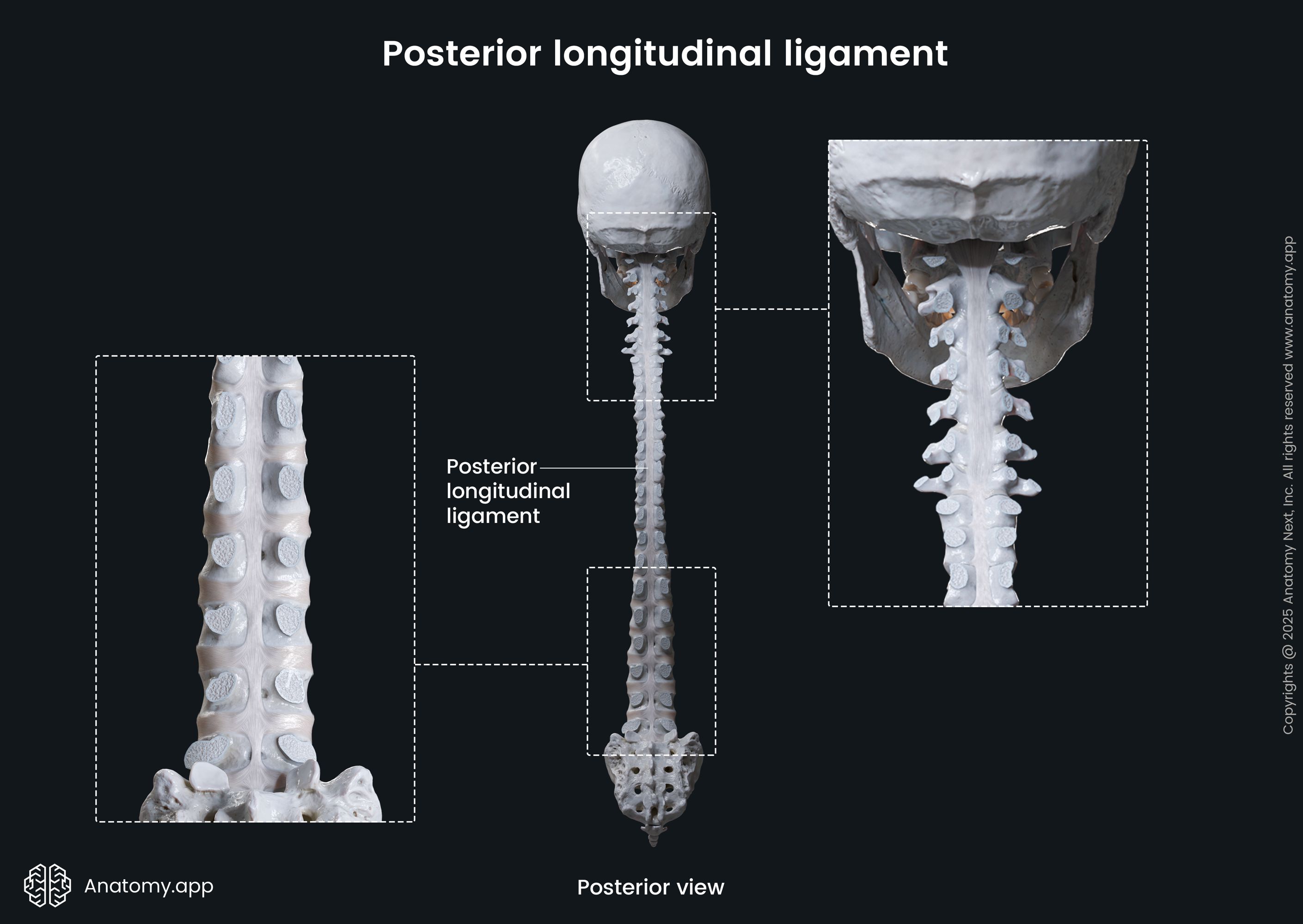
The cervical and thoracic parts of the posterior longitudinal ligament are broader and more uniform in width than the lower parts. In the lower thoracic and lumbar spine, it is narrower over the vertebral bodies and wider over the intervertebral discs. This denticulate appearance is the most characteristic feature of the posterior longitudinal ligament.
The posterior longitudinal ligament has extensions that are called trabeculations. They are made up of connective tissue that binds it to the posteriorly located dura mater - the outermost membrane covering and enclosing the spinal cord. A network of various internal vertebral venous blood vessels of the epidural sinuses (Batson’s plexus) passes between the dura mater and the posterior longitudinal ligament.
Posterior longitudinal ligament composition
The posterior longitudinal ligament is composed of two layers of fibers - superficial and deep. The superficial fibers are longer than the deep and connect three or four vertebral levels. The deep layer of fibers extends only between two adjacent vertebrae and forms extensions that travel through the intervertebral foramina. These fibers firmly adhere to the intervertebral discs. They bond to annuli fibrosi, hyaline cartilages of the end-plates and margins of neighboring vertebral bodies.
The fibers of the posterior longitudinal ligament form a thick band between the vertebral pedicles, especially in the lower thoracic and lumbar regions of the spine. This band of connective tissue is loosely attached to the vertebral bodies and allows the blood vessels to pass through it. Here the basivertebral veins leave the vertebral bodies and enter the anterior internal vertebral venous plexuses.
Posterior longitudinal ligament function
The posterior longitudinal ligament contributes to the strength, balance and firmness of all components of the vertebral column. It secures the posterior aspects of the intervertebral discs, protecting them from herniation. Also, the posterior longitudinal ligament increases the overall stability of the spine during flexion.
References:
- Gray, H., & Carter, H. (2021). Gray’s Anatomy (Leatherbound Classics) (Leatherbound Classic Collection) by F.R.S. Henry Gray (2011) Leather Bound (2010th Edition). Barnes & Noble.
- Paulsen, F., Waschke, J., Hombach-Klonisch, S., Klonisch, T., & Peeler, J. (2019). Sobotta Clinical Atlas of Human Anatomy, one volume, English (1st ed.). Urban & Fischer.
- Steinmetz, M., & Benzel, E. (2016). Benzel’s Spine Surgery, 2-Volume Set: Techniques, Complication Avoidance and Management (4th ed.). Elsevier.
- Watson, C., Paxinos, G., & Kayalioglu, G. (2008). The Spinal Cord: A Christopher and Dana Reeve Foundation Text and Atlas (1st ed.). Academic Press.
Anatomy.app
Contact information
- For questions regarding business inquiries. Please contact:
- info@anatomy.app