- Anatomical terminology
- Skeletal system
- Joints
- Classification of joints
- Joints of skull
- Joints of spine
- Joints of lower limb
- Muscles
- Heart
- Blood vessels
- Lymphatic system
- Nervous system
- Respiratory system
- Digestive system
- Urinary system
- Female reproductive system
- Male reproductive system
- Endocrine glands
- Eye
- Ear
Supraspinous and nuchal ligaments
The supraspinous ligament (Latin: ligamentum supraspinale) is a strong cord-like band that runs along the tips of the vertebral spinous processes beginning from the 7th cervical vertebra (C7) and terminating at the lower lumbar vertebrae and the sacrum. Most commonly, it ends at the fourth lumbar vertebra (L4) level.
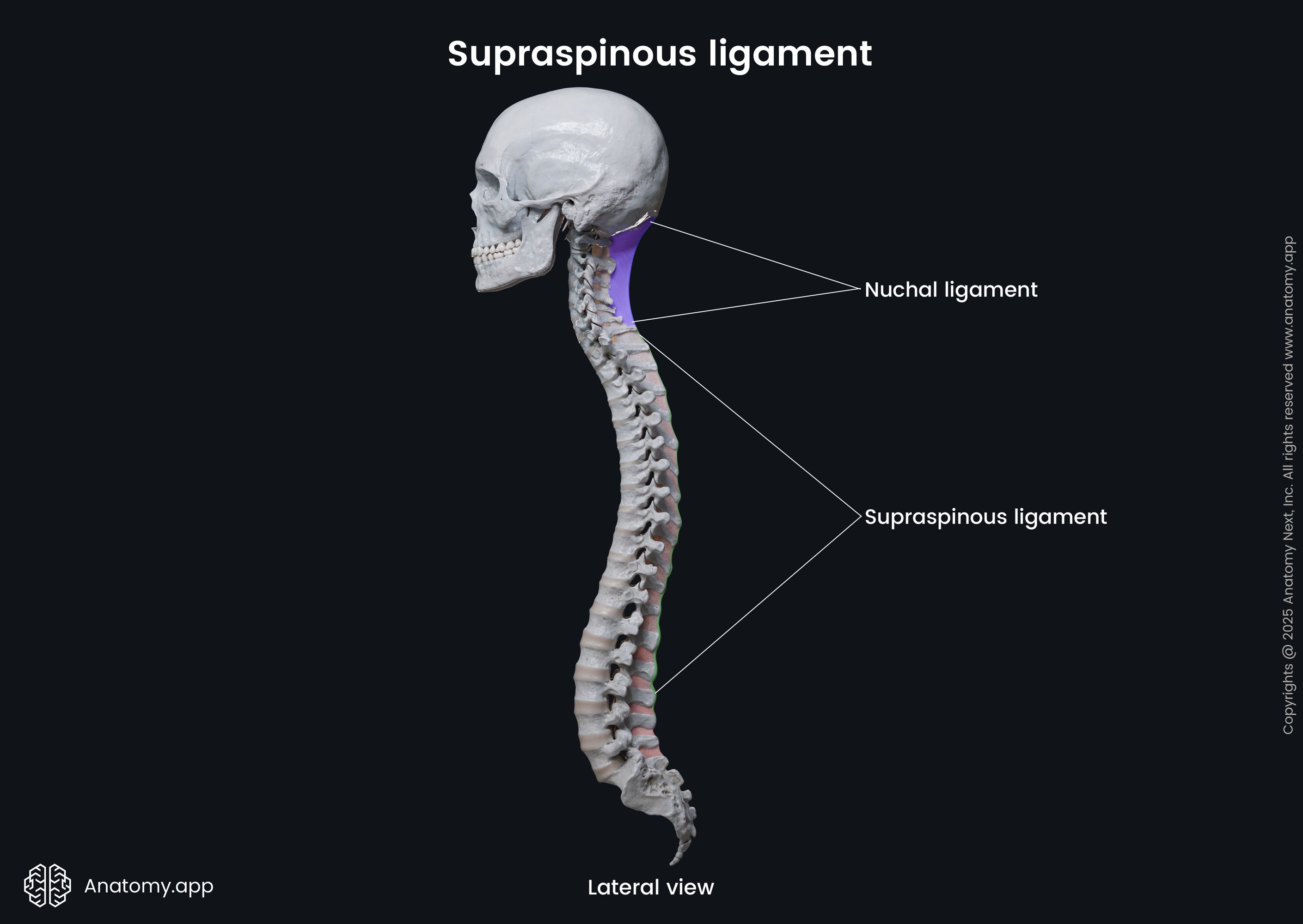
Starting from the C7 vertebrae, the supraspinous ligament continues as the nuchal ligament (Latin: ligamentum nuchae). It extends in the cranial direction to reach the external occipital protuberance, where it terminates. Structurally the nuchal ligament differs from the upper part of the supraspinous ligament, and it is rather referred to as a triangular fibrous membrane.
Supraspinous ligament anatomy
The supraspinous ligament is a strong fibrous cord extending along the thoracic and lumbar spine. It has superficial, middle and deep layers of fibers. The superficial fibers span three to four spinal vertebrae, and the middle layer of fibers connects two to three vertebrae. And finally, the deep layer links adjacent spinous processes. Anteriorly, the supraspinous ligament fuses with the interspinous ligaments. The middle and deep layers mainly consist of fibers from the aponeurosis of the longissimus thoracis muscle.
Nuchal ligament anatomy
The nuchal ligament is a triangular-shaped fibrous structure composed of fascia and tendons. It is oriented in the sagittal plane and found between the posterior muscles of the neck along the midline of the cervical spine. The nuchal ligament has a base and an apex. The apex attaches to the tip of the spinous process of the C7 vertebra. Inferiorly, it is continuous with the supraspinous ligament.
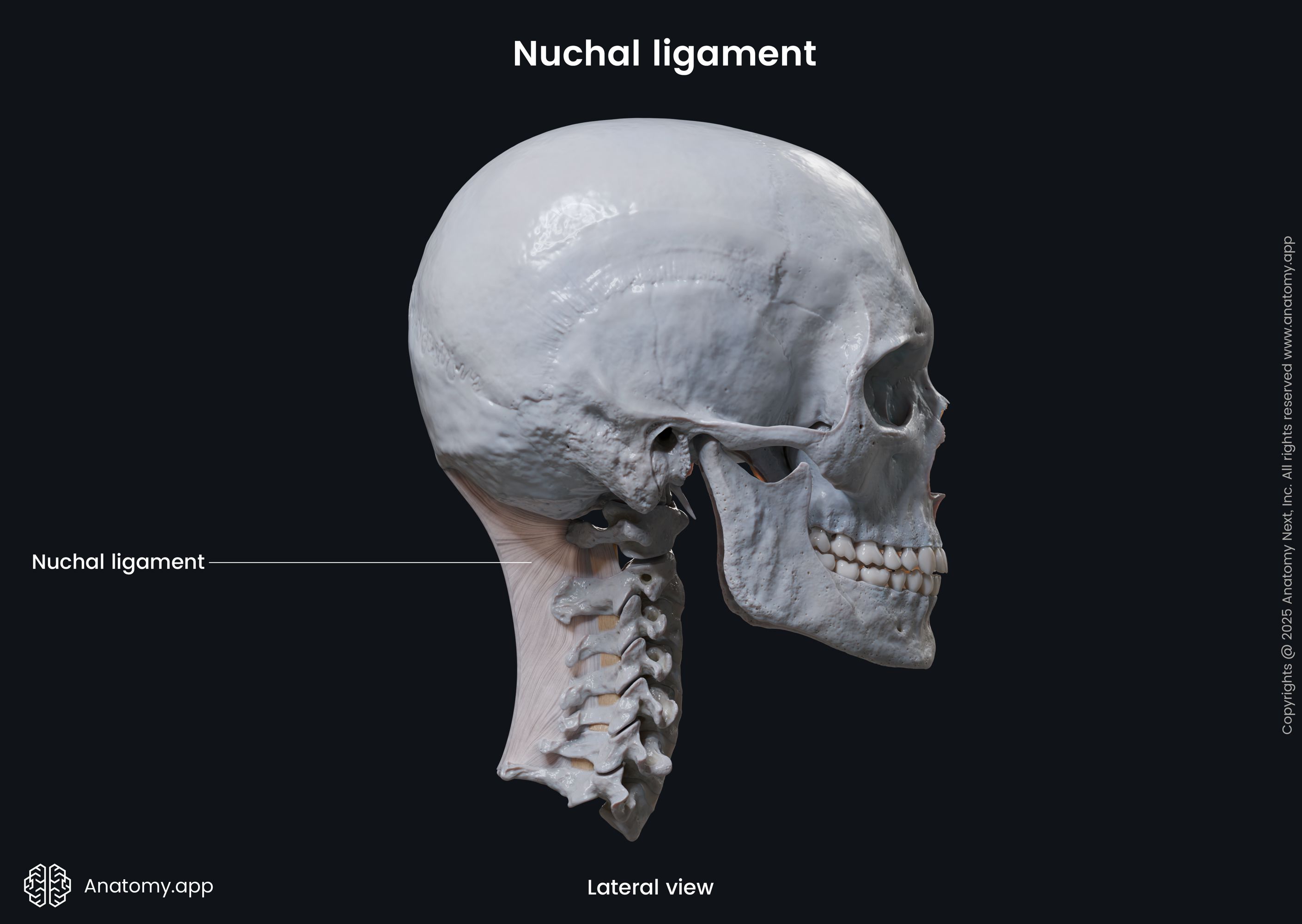
The base of the nuchal ligament attaches to the skull - it adheres to the external occipital protuberance and goes down to the foramen magnum. One margin of the triangular ligament crosses over the spinous processes of the cervical vertebrae and the posterior tubercle of the first cervical vertebra (C1, atlas), while the other margin is free.
The nuchal ligament can be divided into two parts - the dorsal raphe and median (midline) fascial septum. The dorsal raphe lies superficially in the posterior midline of the neck. Therefore, it extends between the C7 spinous process and the external occipital protuberance. The superior part of the dorsal raphe is formed by the interweaving of the right and left cervical parts of the trapezius muscles. Inferiorly, the dorsal raphe consists of tendons from the splenius capitis and rhomboid minor muscles.
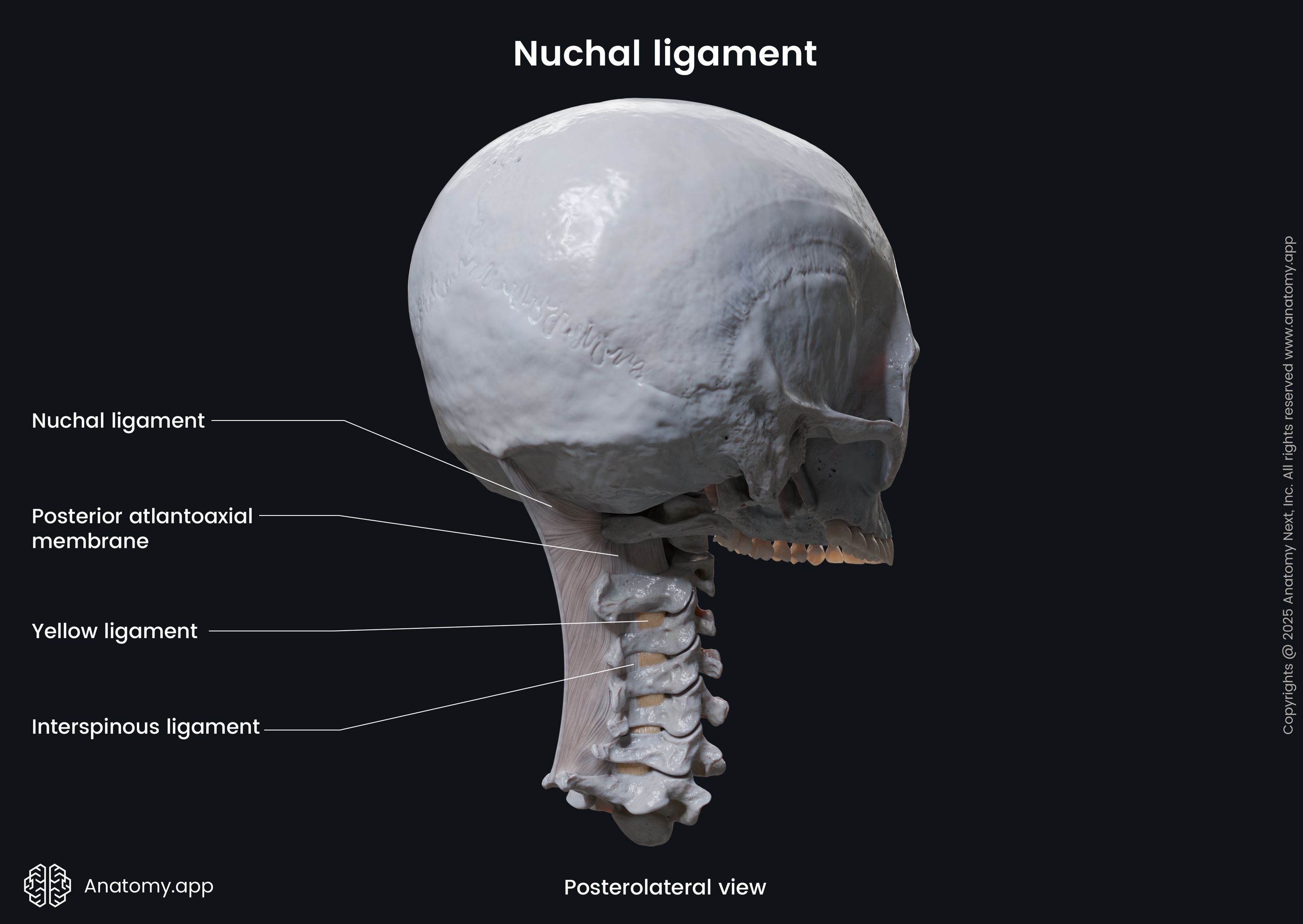
The anterior portion of the dorsal raphe that extends towards the spine forms the median fascial septum. It completely separates the semispinalis capitis muscle from the opposite semispinalis capitis, as well as from the multifidus and semispinalis cervicis muscles. Anteriorly, the median fascial septum blends with the interspinous ligaments, posterior atlanto-occipital and atlanto-axial membranes and is also attached to the posterior aspect of the spinal dura mater.
Functions of supraspinous and nuchal ligaments
The supraspinous ligament provides tension for the entire spine. It prevents hyperflexion of the spine and provides resistance to the separation of the spinous processes. This ligament also serves as an attachment site for muscles that are located along the midline of the spine, such as the trapezius.
The nuchal ligament limits flexion of the head and cervical spine and helps to return the head to its anatomical position. Therefore, the nuchal ligament supports the head. It also serves as an attachment site for adjacent muscles of the neck and upper back.
References:
- Drake, R., Vogl, W., & Mitchell, A. (2019). Gray’s Anatomy for Students: With Student Consult Online Access (4th ed.). Elsevier.
- Gray, H., & Carter, H. (2021). Gray’s Anatomy (Leatherbound Classics) (Leatherbound Classic Collection) by F.R.S. Henry Gray (2011) Leather Bound (2010th Edition). Barnes & Noble.
- Steinmetz, M., & Benzel, E. (2016b). Benzel’s Spine Surgery, 2-Volume Set: Techniques, Complication Avoidance and Management (4th ed.). Elsevier.
Anatomy.app
Contact information
- For questions regarding business inquiries. Please contact:
- info@anatomy.app