- Anatomical terminology
- Skeletal system
- Joints
- Classification of joints
- Joints of skull
- Joints of spine
- Joints of lower limb
- Joints of pelvis
- Hip joint
- Knee joint
- Tibiofibular joints
- Joints of foot
- Muscles
- Heart
- Blood vessels
- Lymphatic system
- Nervous system
- Respiratory system
- Digestive system
- Urinary system
- Female reproductive system
- Male reproductive system
- Endocrine glands
- Eye
- Ear
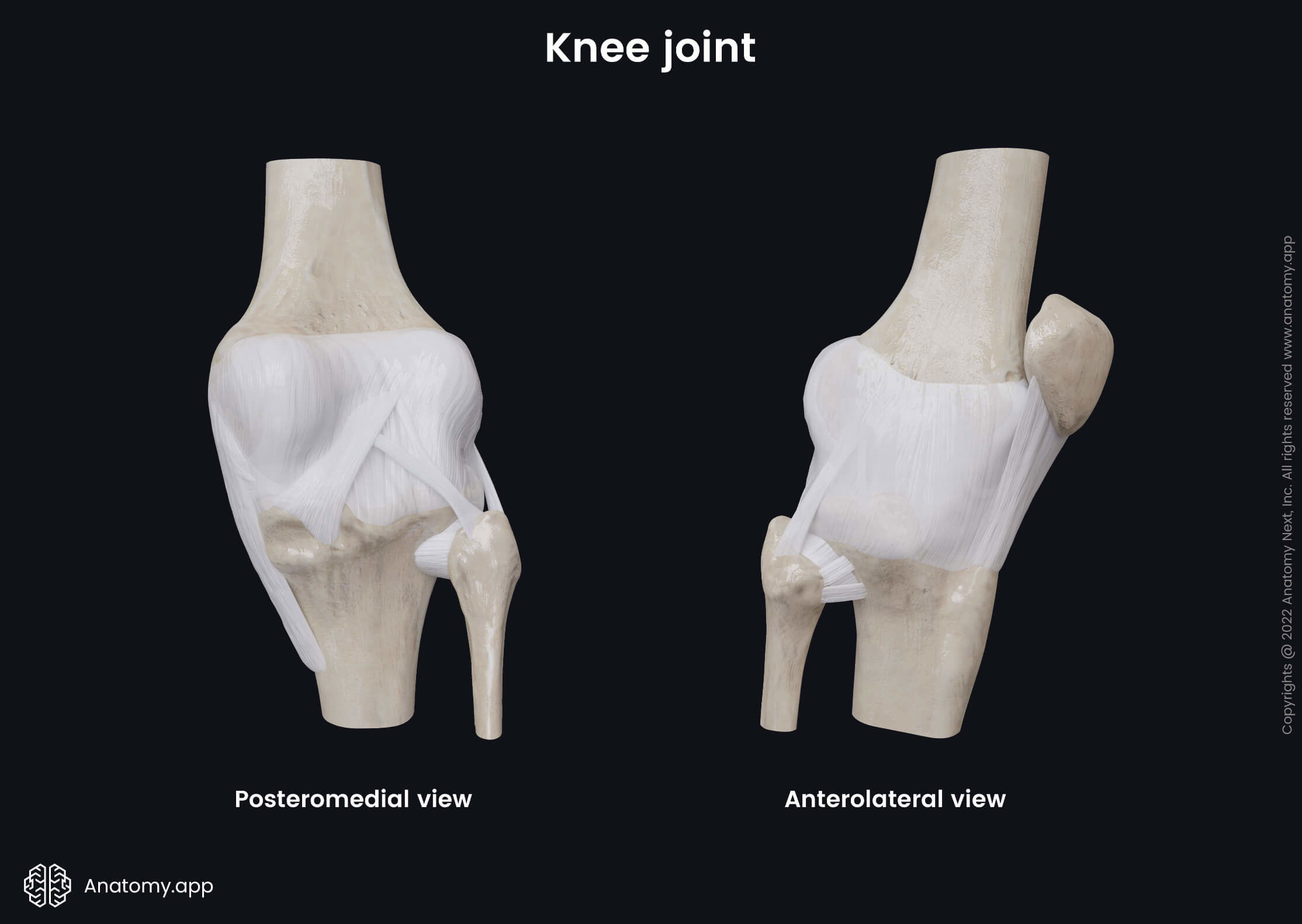
Knee joint
The knee joint (Latin: articulatio genus) is a synovial hinge type joint that is formed between three bones - femur, tibia and patella. The knee joint is the most complicated and one of the strongest joints in the human body. Besides the joint capsule and ligaments that strengthen it, this articulation also contains menisci and bursae.
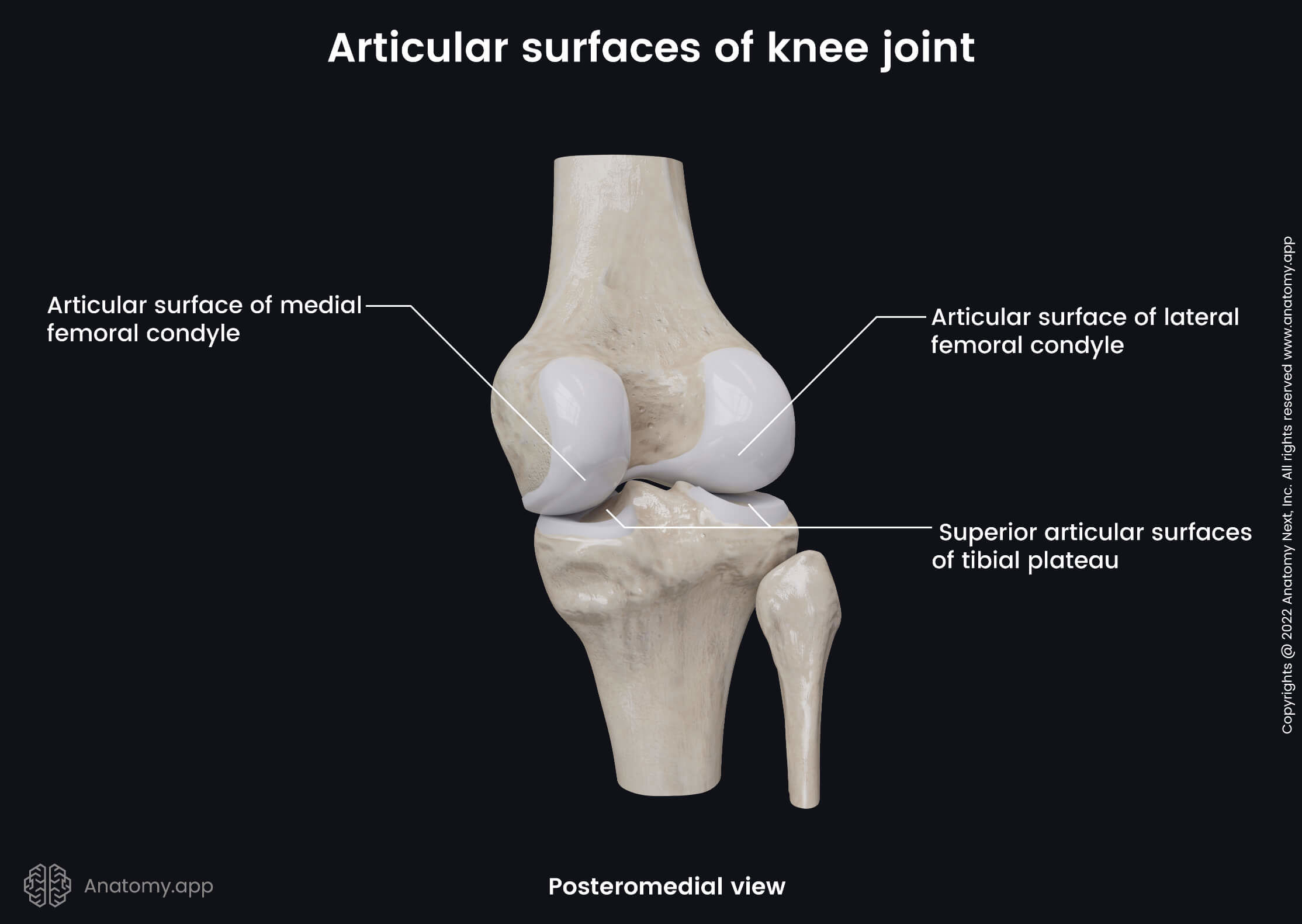
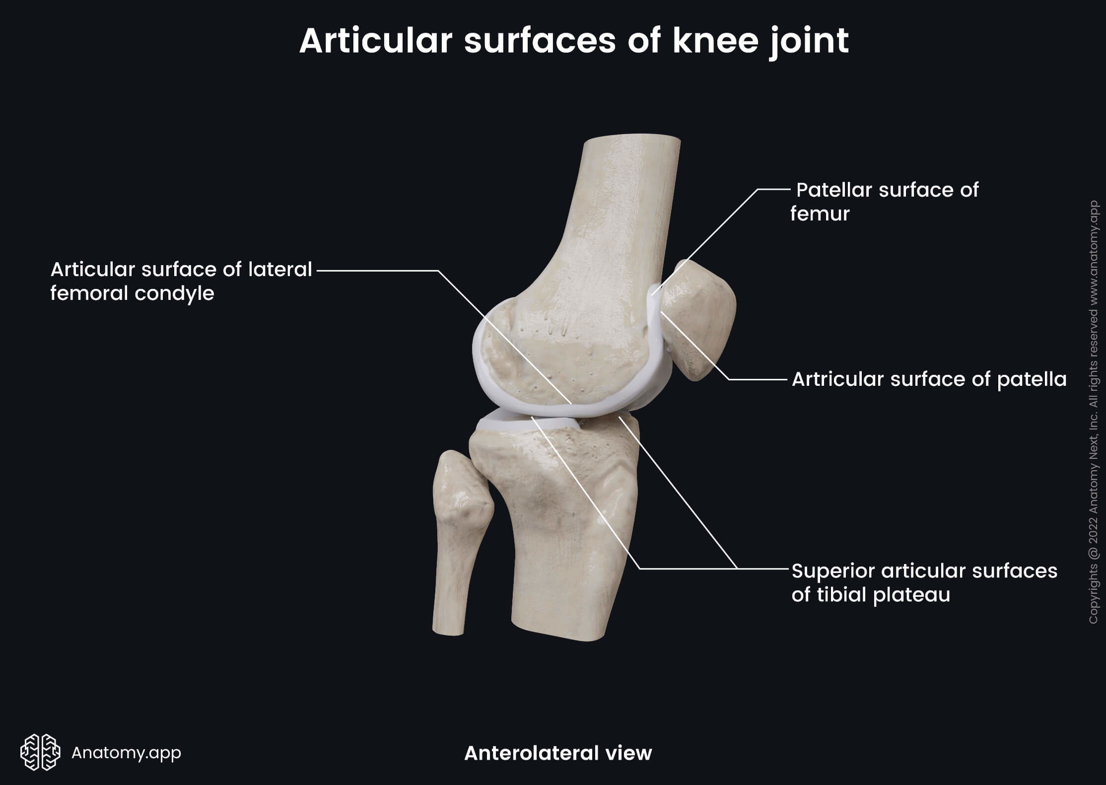
Articulating structures of knee joint
The knee joint is composed of two articulations - the tibiofemoral joint between the tibia and femur and the patellofemoral articulation between the patella and femur. Structures that articulate in the knee joint are as follows:
- Articular surfaces of the medial and lateral condyles of the femur
- Superior articular surfaces of the tibia (tibial plateau)
- Patellar surface of the femur
- Articular surface of the patella
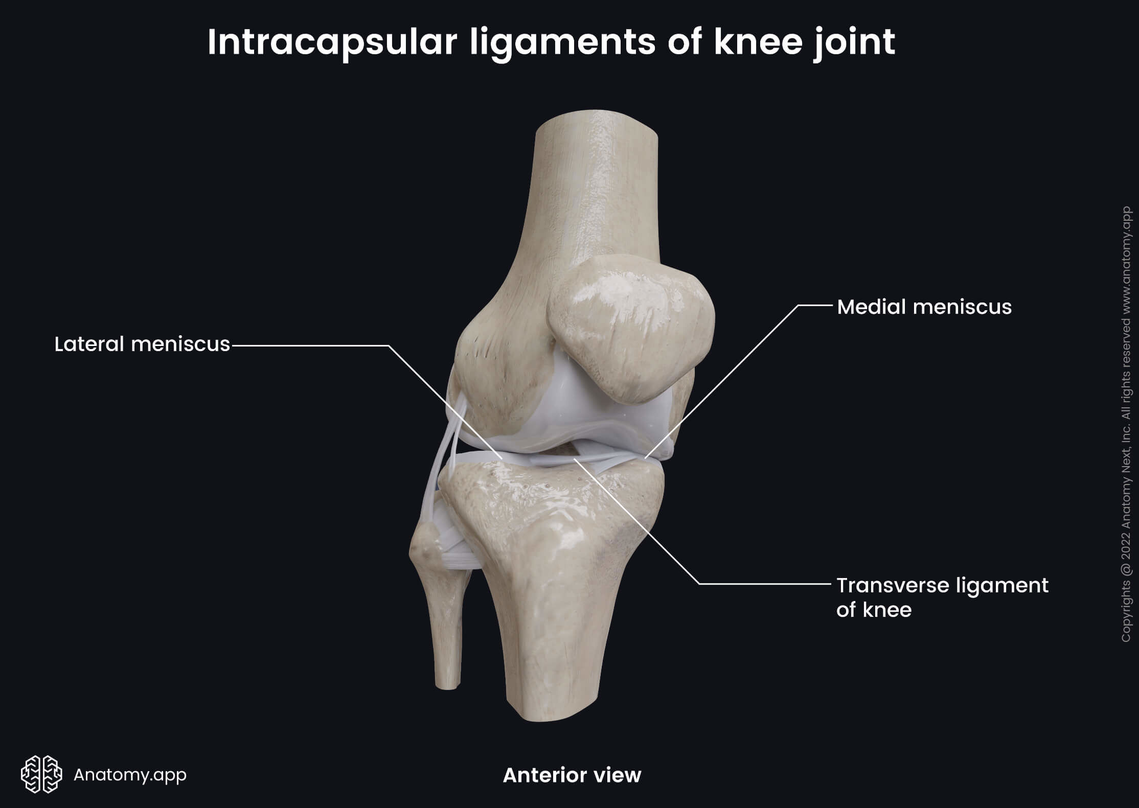
Menisci
Between the surfaces of the tibiofemoral joint are located thin C-shaped fibrocartilage plates known as the menisci. Each knee joint contains two menisci - the medial meniscus and lateral meniscus. The medial meniscus is crescent-shaped, and it appears almost semicircular. In contrast, the lateral meniscus looks like an incomplete circle as it is nearly circular.
The outer parts of both menisci are thicker and contain blood vessels, while the inner aspects are thin and do not contain any blood vessels. The inferior surfaces of menisci appear flat, while the superior ones are concave. The menisci provide a smooth surface and smooth out discrepancies. They protect the bones from damage and improve the stability of the knee joint.
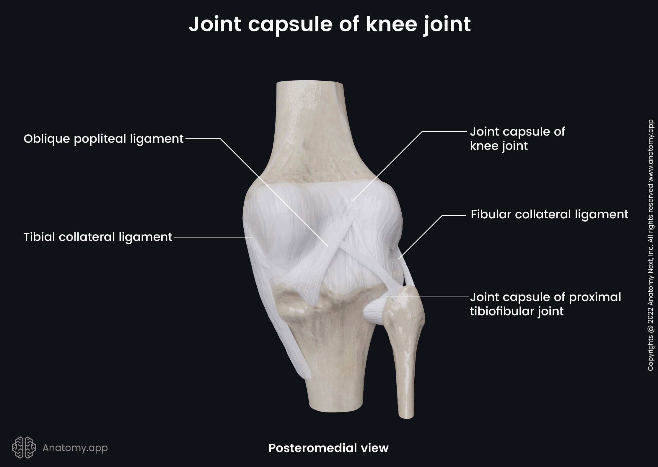
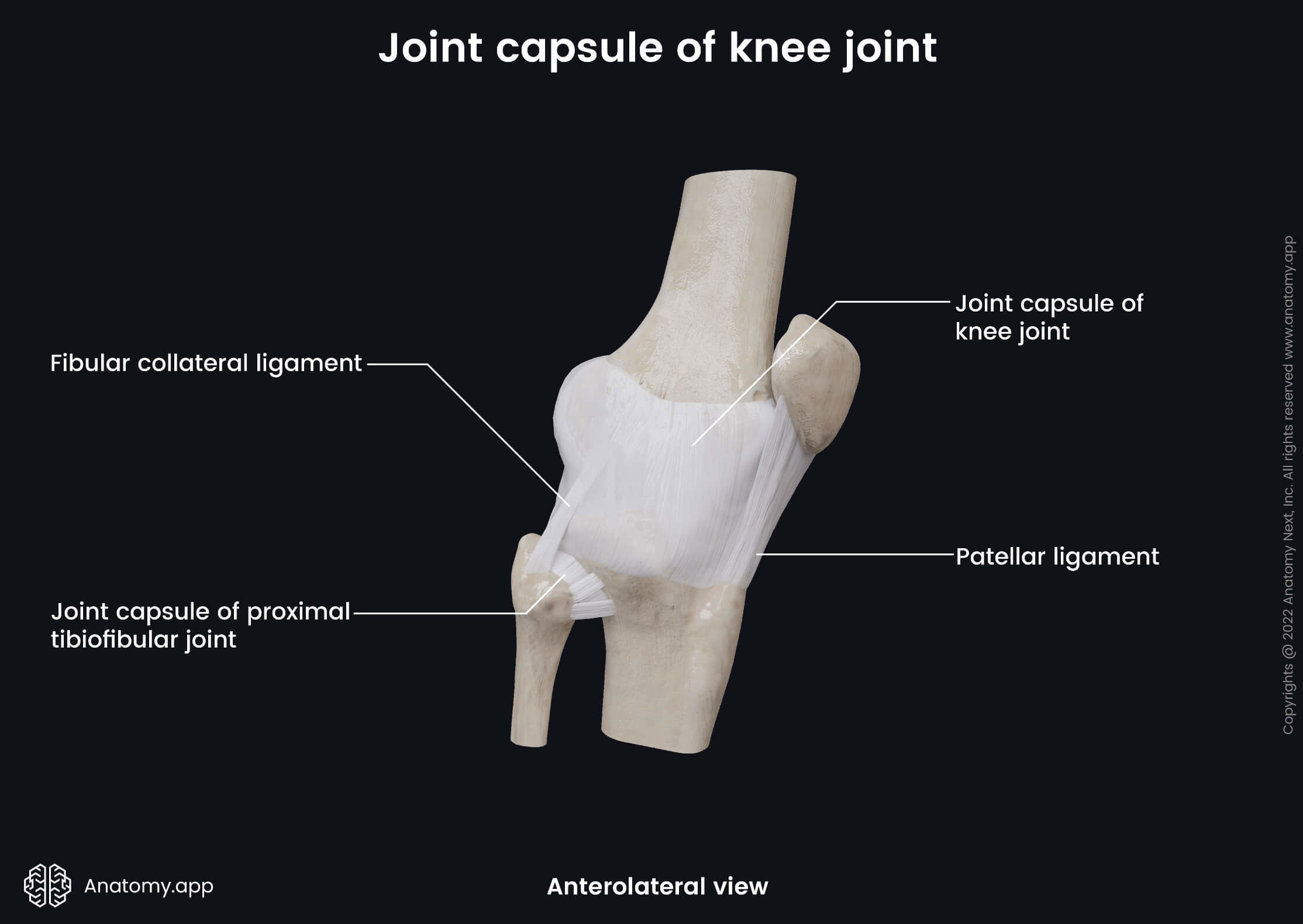
Joint capsule
The knee joint is surrounded by a fibrous joint capsule that at the front of the femur is attached to the edges of the joint surfaces. On the sides of the femur, it also passes along the edges of the joint surfaces. While at the back of the bone, it runs along the upper edge of the intercondylar fossa. For the patella and tibia, the joint capsule attaches along the edges of the joint surfaces.
Bursae
The synovial membrane of the joint capsule fills the hollow spaces between the bones, and it produces an oily synovial fluid that lubricates the joint to reduce friction. The synovial membrane creates several formations called bursae around the knee joint.
A bursa is a fluid-filled synovial sac between moving structures in a joint. Four bursae within the knee joint communicate with the synovial cavity, and they include the following:
- Anserine bursa - separates the tendon of the sartorius, gracilis and semitendinosus muscles from the tibia and the tibial collateral ligament;
- Suprapatellar bursa - situated between the femur and the tendon of the quadriceps femoris muscle;
- Semimembranosus-gastrocnemius bursa - positioned anteriorly to the tendon of the medial head of the gastrocnemius;
- Popliteus bursa - located between the tendon of the popliteus muscle and the lateral condyle of the tibia.
Besides bursae connected with the synovial cavity, the knee joint contains several more bursae that are not connected to it. These bursae are as follows:
- Subcutaneous prepatellar bursa - located between the skin and anterior surface of the patella;
- Subcutaneous infrapatellar bursa - situated between the skin and the tibial tuberosity;
- Deep infrapatellar bursa - between the patellar ligament and the anterior surface of the tibia; sometimes this bursa is connected with the synovial cavity.
Ligaments of knee joint
The stability of the knee joint is increased with the help of fibrous connective tissue bands called ligaments. They help to hold the joint in place. The knee joint is strengthened by two groups of ligaments - the intracapsular and extracapsular ligaments.
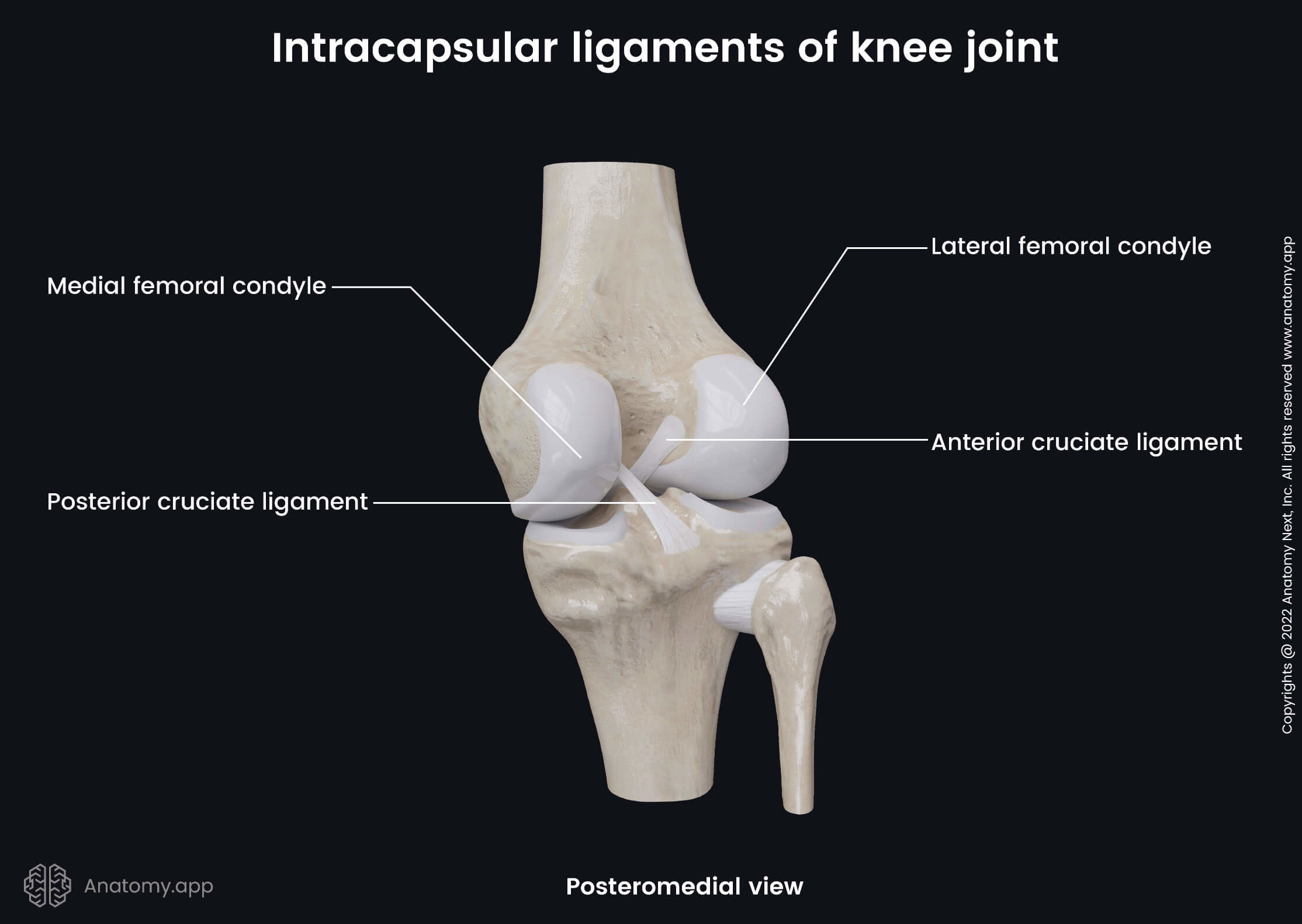
Intracapsular ligaments
The intracapsular ligaments are located inside the knee joint cavity, and they include three ligaments:
- Transverse ligament of the knee - extends anteriorly between both menisci;
- Anterior cruciate ligament - stretches from the inner surface of the lateral condyle of the femur to the anterior intercondylar area of the tibia;
- Posterior cruciate ligament - extends from the lateral surface of the medial condyle of the femur to the posterior intercondylar area of the tibia.
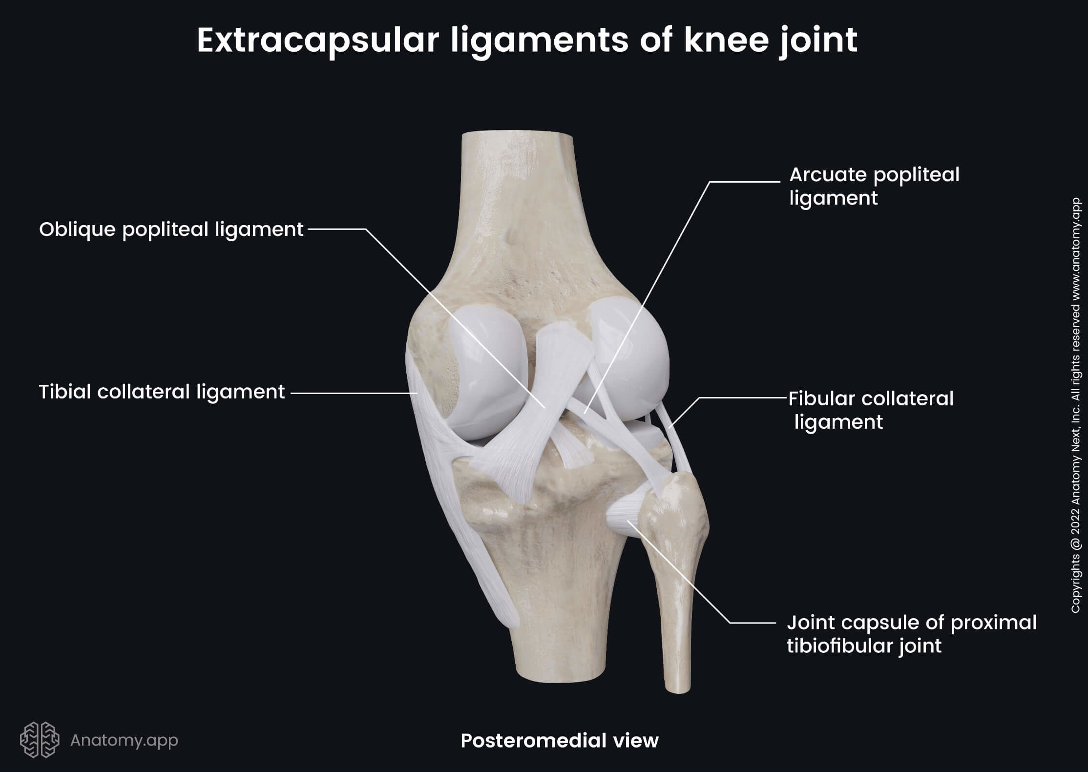
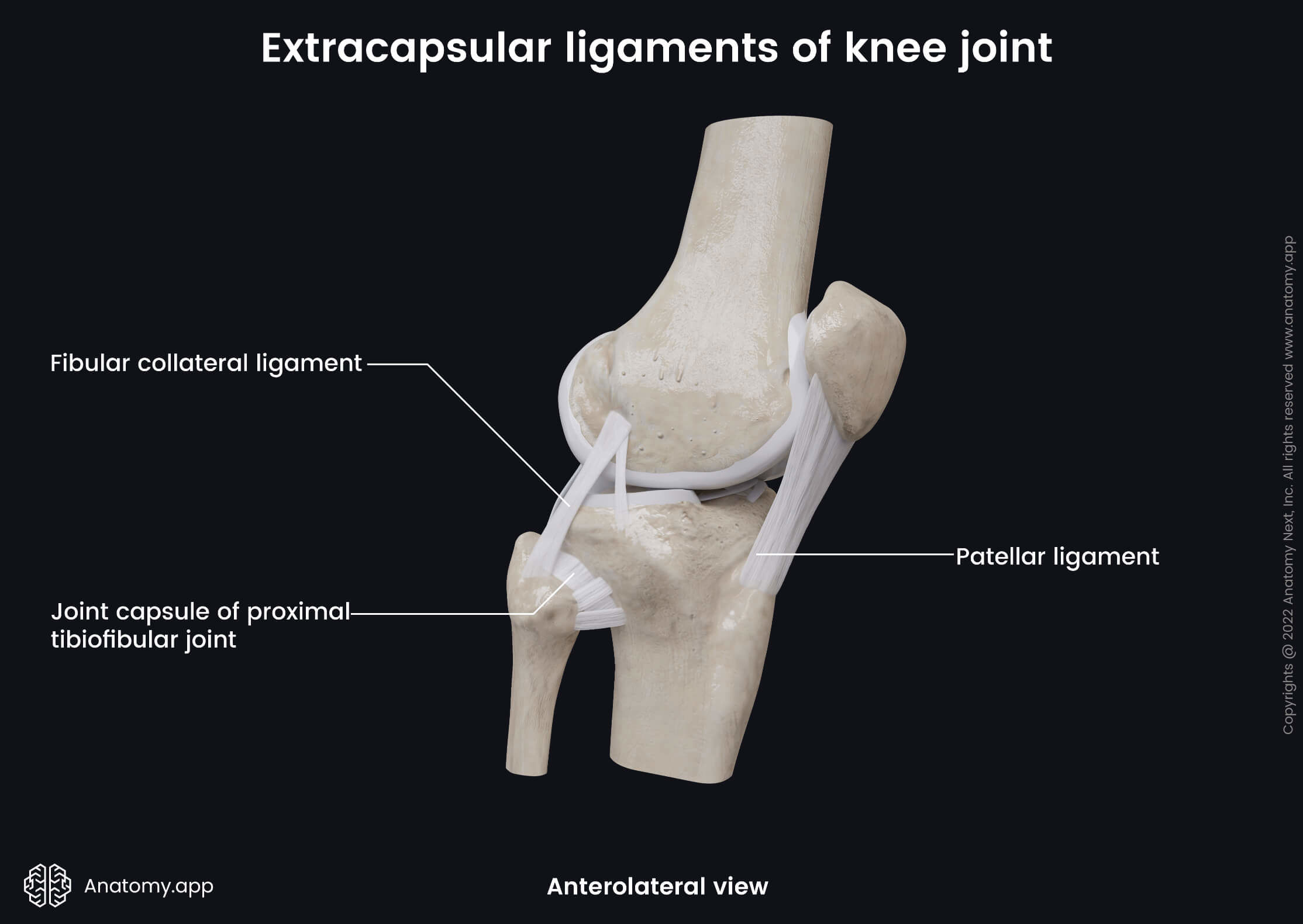
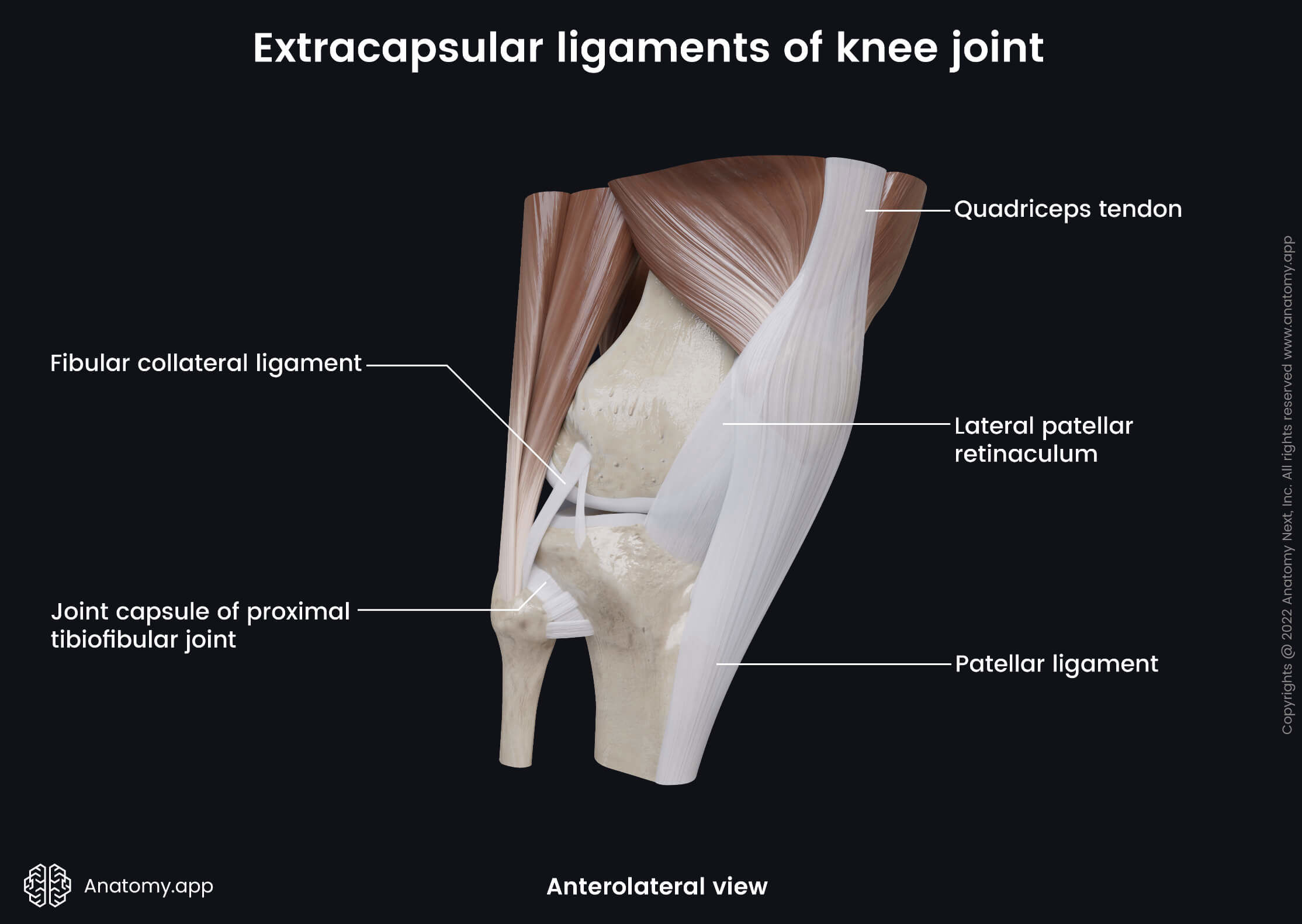
Extracapsular ligaments
The extracapsular ligaments are located outside the capsule and are continuous with its outer surface. The knee joint contains seven extracapsular ligaments:
- Tibial collateral ligament - passes from the medial epicondyle of the femur to the medial condyle and surface of the body of the tibia;
- Fibular collateral ligament - stretches from the lateral epicondyle of the femur to the lateral side of the head of the fibula;
- Oblique popliteal ligament - located on the posterior side of the joint; extends from the lateral condyle of the femur to the medial condyle of the tibia;
- Arcuate popliteal ligament - situated on the posterior side of the joint; it stretches from the head of the fibula to the oblique popliteal ligament;
- Medial patellar retinaculum - made up of the fibers of the vastus medialis;
- Lateral patellar retinaculum - formed by the fibers of the vastus lateralis;
- Patellar ligament - a continuation of the tendon of the quadriceps femoris muscle.
Movements of knee joint
The knee joint permits four movements:
- Flexion and extension of the leg
- External (lateral) and internal (medial) rotation of the leg
Anatomy.app
Contact information
- For questions regarding business inquiries. Please contact:
- info@anatomy.app