- Anatomical terminology
- Skeletal system
- Joints
- Classification of joints
- Joints of skull
- Joints of spine
- Joints of lower limb
- Joints of pelvis
- Hip joint
- Knee joint
- Tibiofibular joints
- Joints of foot
- Muscles
- Heart
- Blood vessels
- Lymphatic system
- Nervous system
- Respiratory system
- Digestive system
- Urinary system
- Female reproductive system
- Male reproductive system
- Endocrine glands
- Eye
- Ear
Hip joint
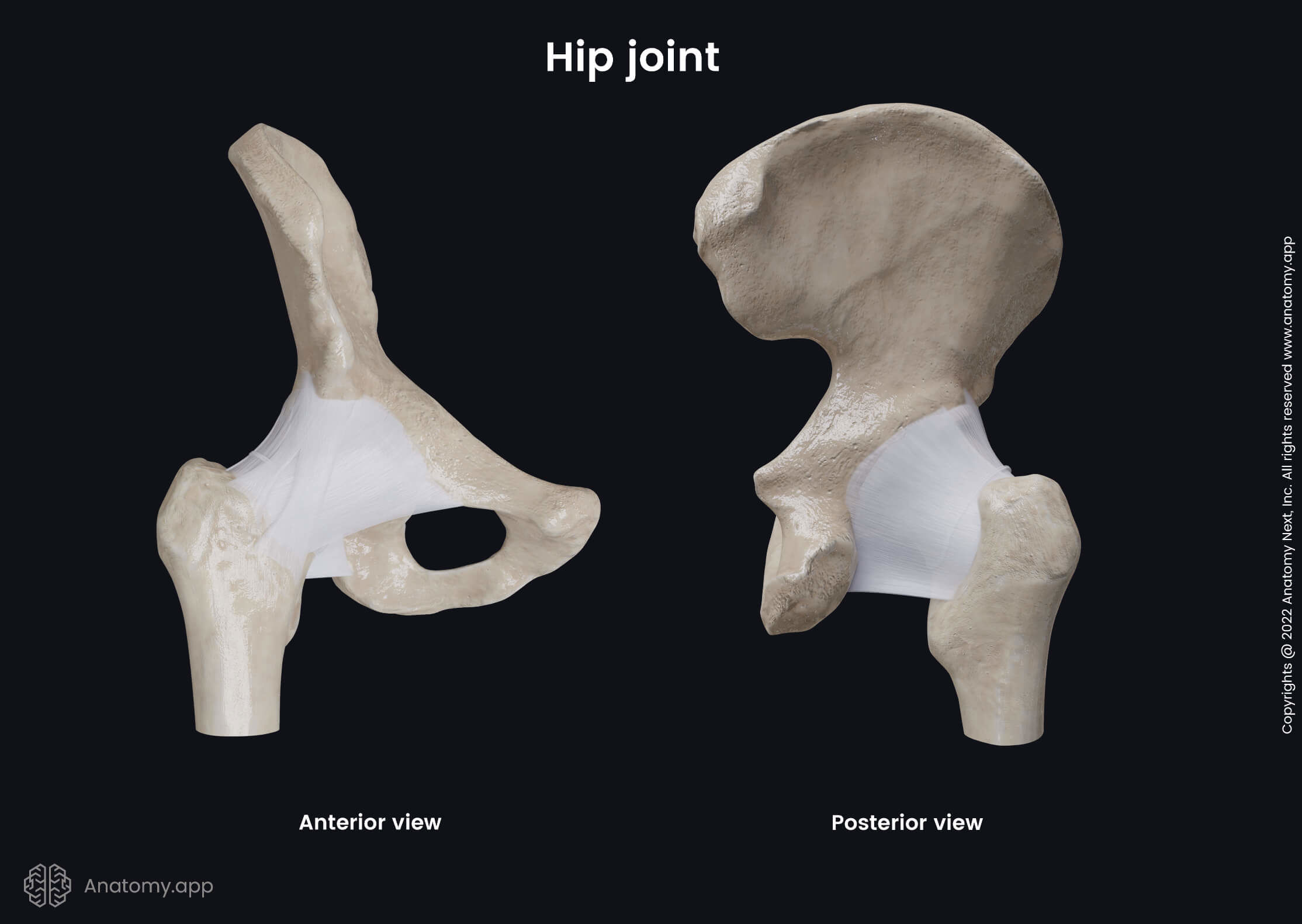
The hip joint (Latin: articulatio coxae), also known as the coxofemoral or acetabulofemoral joint, is an articulation formed between the acetabulum of the pelvis and the head of the femur. Therefore, the hip joint connects the pelvic girdle to the lower free extremity. It is classified as the synovial ball and socket type joint.
Articulating structures of hip joint
The articulating surfaces of the hip joint are as follows:
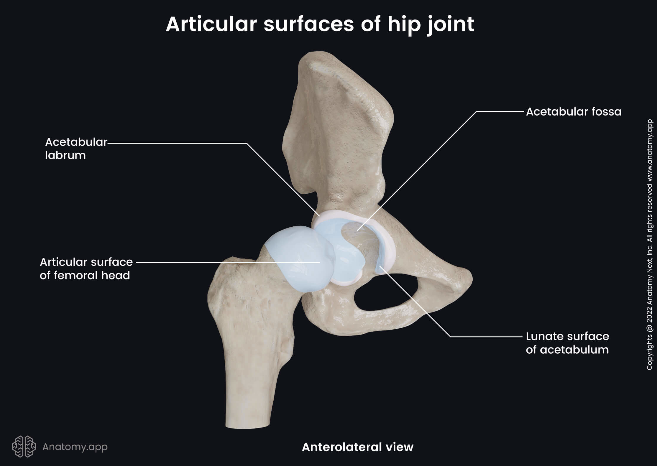
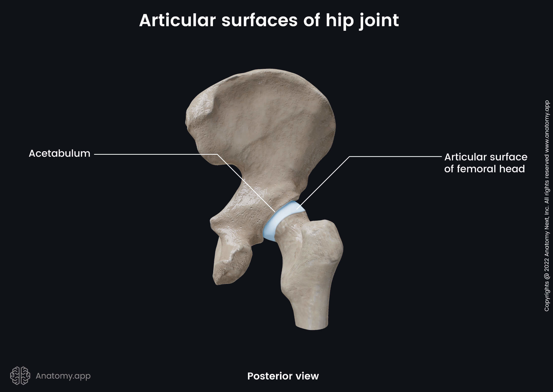
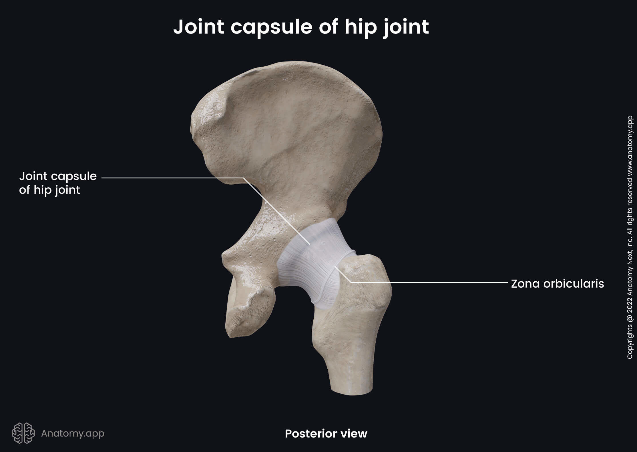
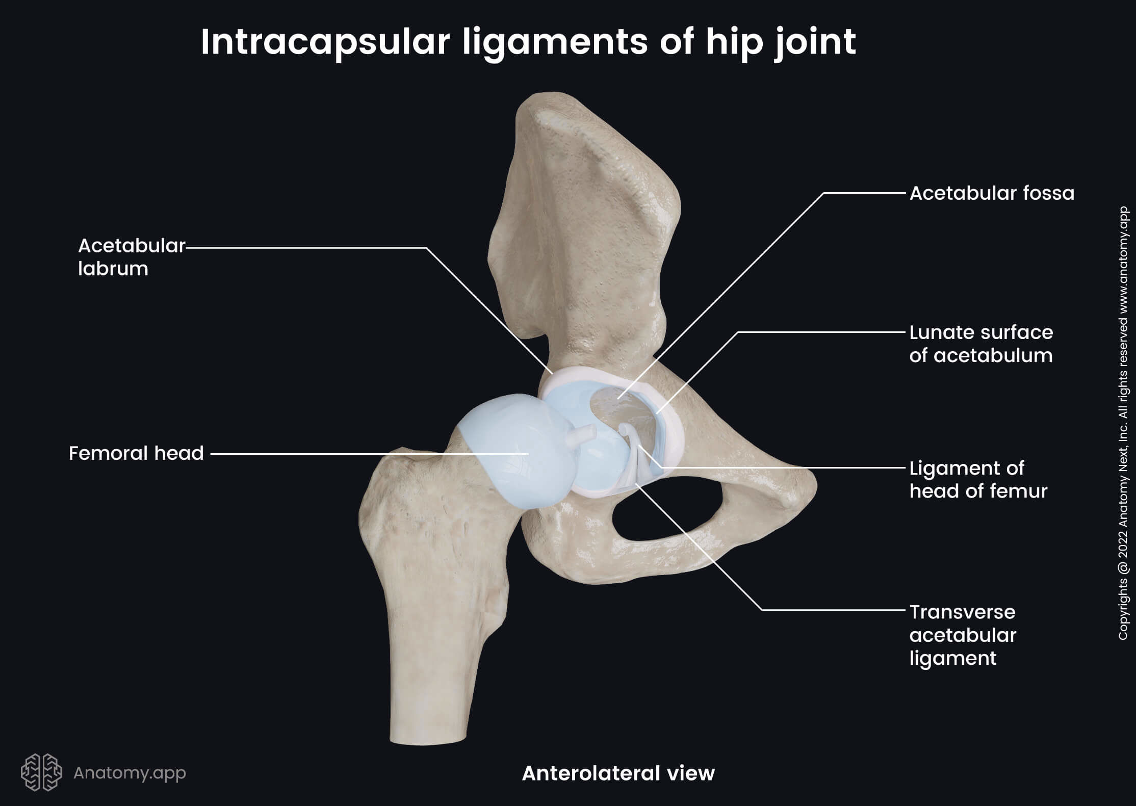
The acetabulum is a cup-like depression on the inferolateral aspect of the pelvis. It is surrounded by a fibrocartilaginous collar - the acetabular lip or acetabular labrum - that is attached to the external margins of the acetabulum. Both articulating structures are enclosed in a fibrous joint capsule that is attached to the outer edge of the acetabulum and acetabular labrum.
Ligaments of hip joint
The stability of the hip joint is increased with the help of two ligament groups. They are named based on their location in relation to the fibrous capsule - intracapsular and extracapsular ligaments.
The intracapsular ligaments are located inside the hip joint cavity. In contrast, the extracapsular ligaments are situated outside the capsule and are continuous with the outer surface of it.
Intracapsular ligaments
The hip joint cavity contains two intracapsular ligaments. They are known as follows:
- Transverse acetabular ligament - connects the ends of the lunate surface and passes above the acetabular notch;
- Ligament of the head of the femur - a small structure that runs from the acetabular notch and transverse acetabular ligament to the fovea capitis of the femoral head; through the ligament goes the artery to the head of the femur.
Extracapsular ligaments
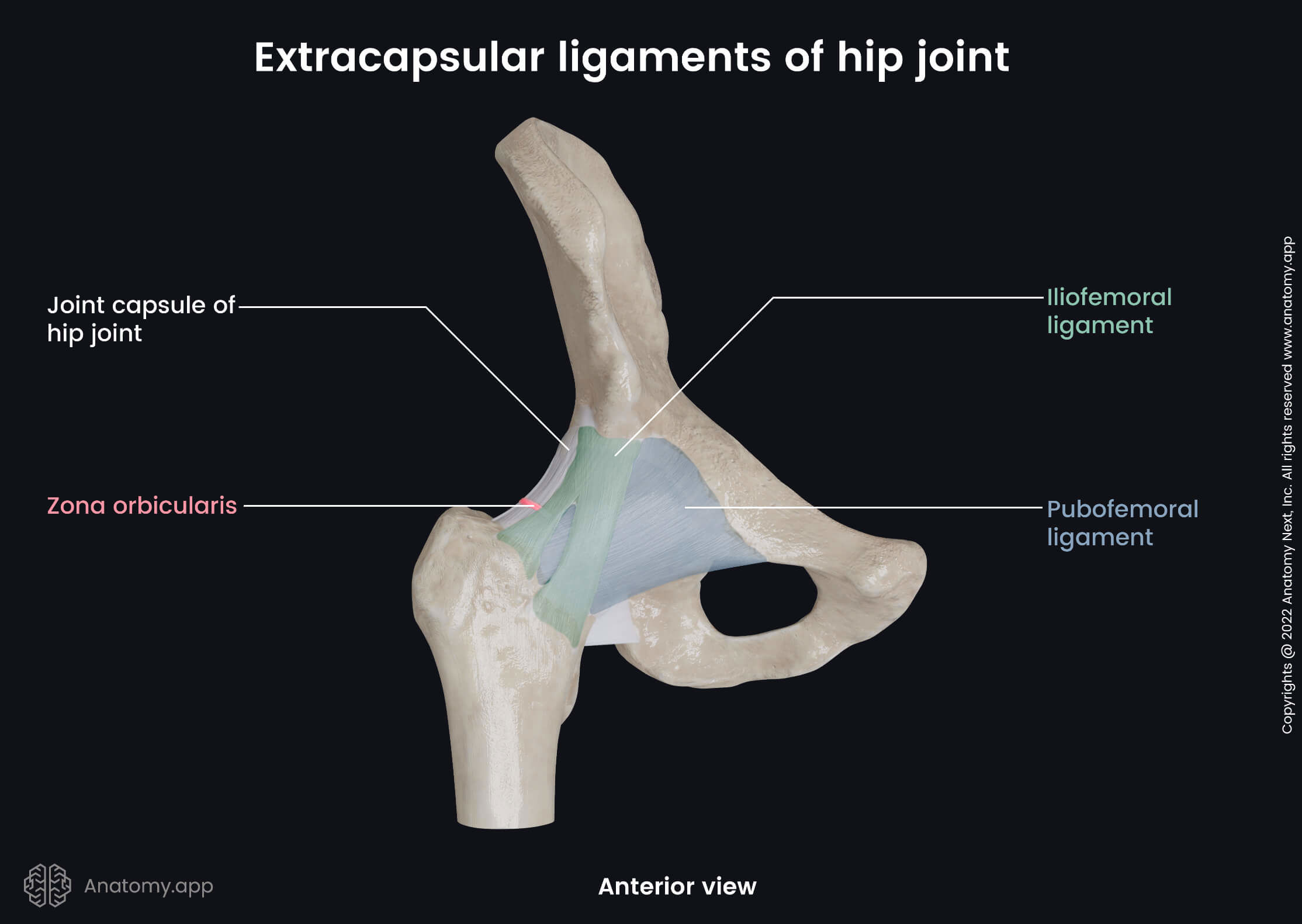
The extracapsular ligaments include four strong bands. These ligaments are as follows:
- Iliofemoral ligament - extends between the anterior inferior iliac spine and the intertrochanteric line of the femur; it is the strongest ligament in the human body; it prevents external rotation and hyperextension of the hip joint;
- Pubofemoral ligament - triangular-shaped ligament located between the superior pubic ramus and the intertrochanteric line of the femur; it prevents the excessive abduction and inner rotation of the thigh at the hip joint;
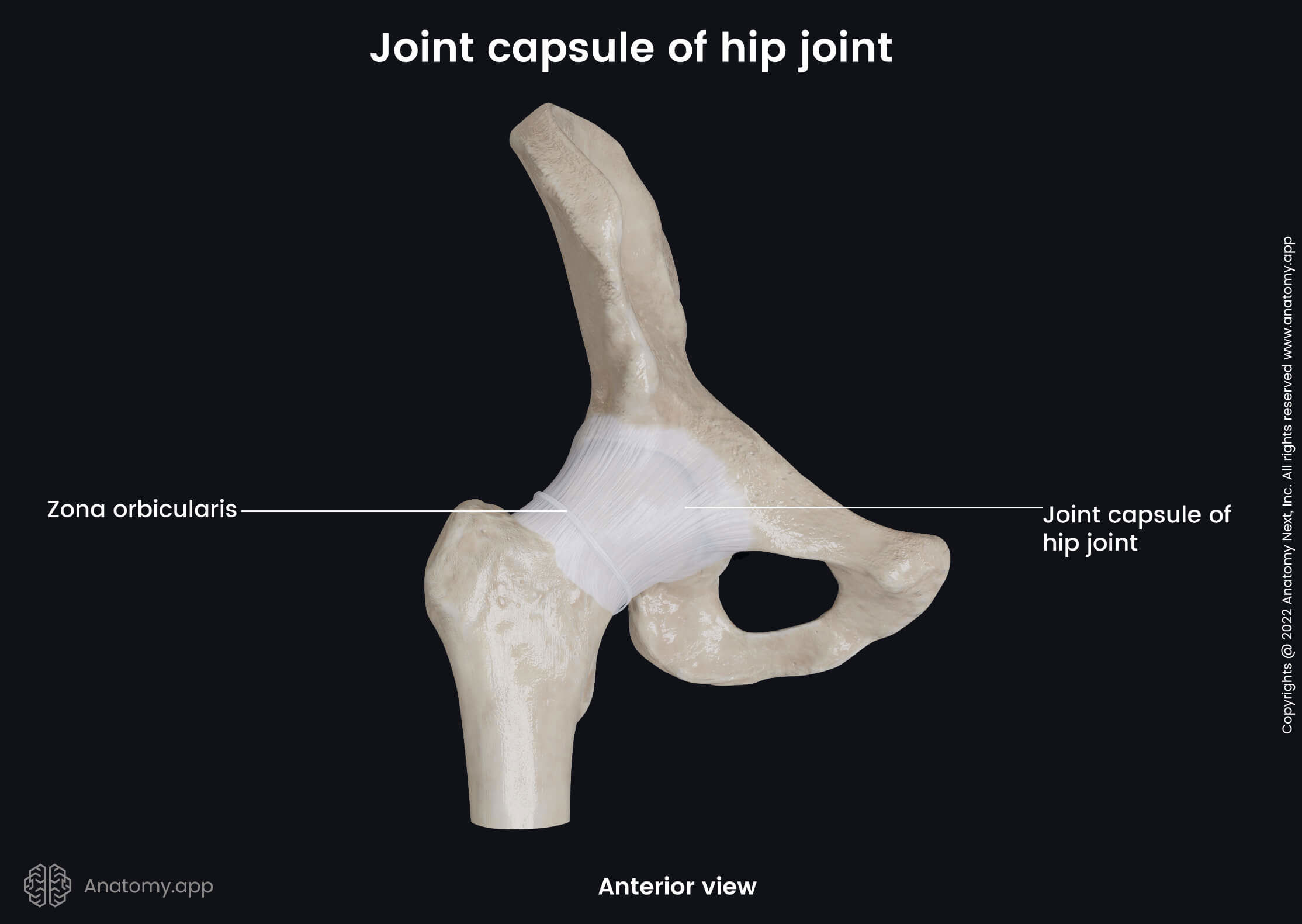
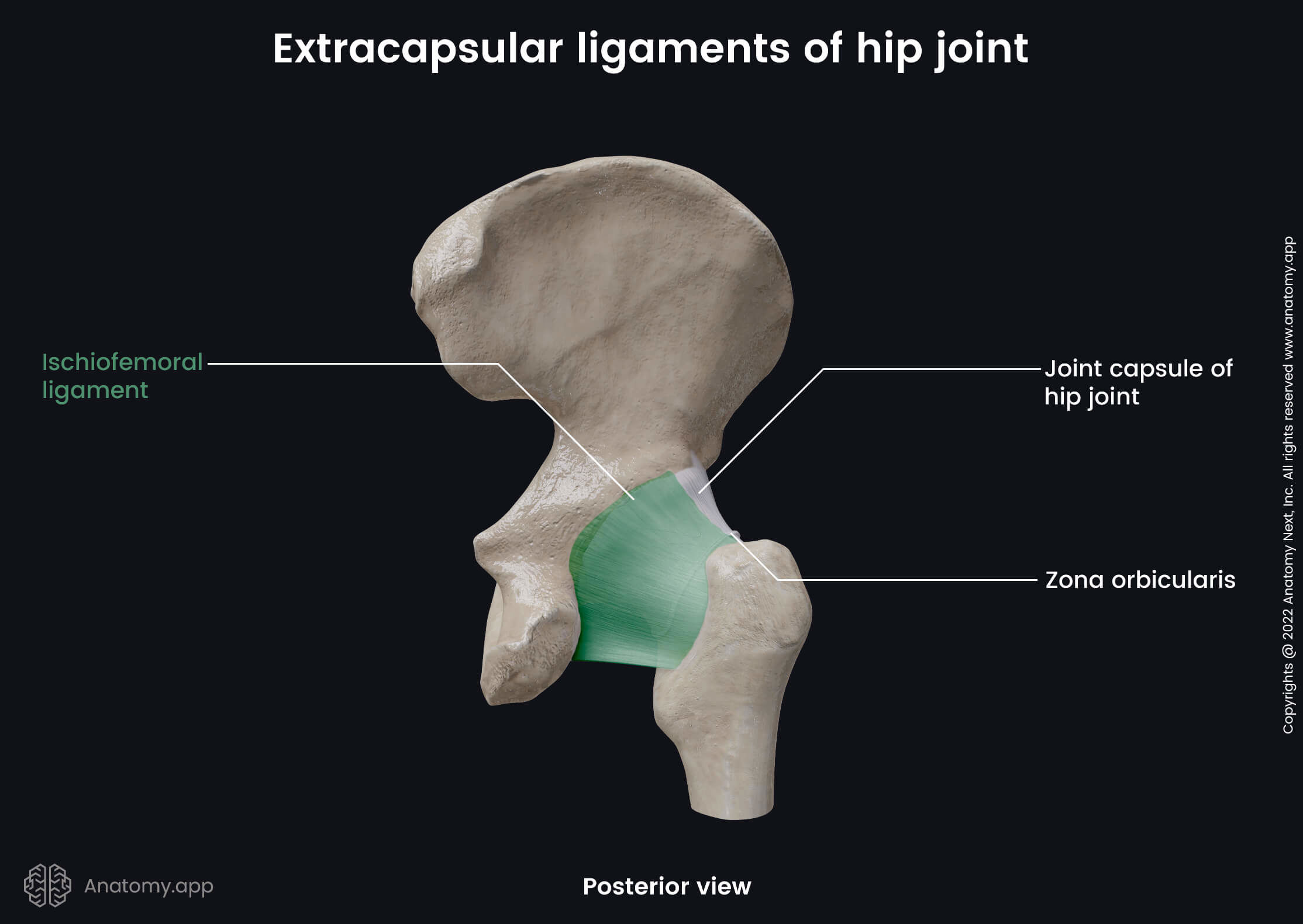
- Zona orbicularis - extends from the anterior inferior iliac spine, surrounds the neck of the femur and ends where it started; it is formed by the circular fibers of the hip joint capsule;
- Ischiofemoral ligament - stretches from the body of the ischium to the trochanteric fossa; it prevents the thigh from excessive inner rotation and adduction at the hip joint.
Movements of hip joint
The hip joint is classified as a multiaxial joint, allowing a wide range of motions. However, the movements can be performed with a limited amplitude as this joint is the most important weight-bearing joint and it is more designed to provide stability to the body. The movements provided by the joint are as follows:
- Flexion and extension of the thigh
- Abduction and adduction of the thigh
- External or lateral rotation of the thigh
- Internal or medial rotation of the thigh
- Circumduction of the thigh
Anatomy.app
Contact information
- For questions regarding business inquiries. Please contact:
- info@anatomy.app