- Anatomical terminology
- Skeletal system
- Skeleton of trunk
- Skull
- Skeleton of upper limb
- Skeleton of lower limb
- Joints
- Muscles
- Heart
- Blood vessels
- Lymphatic system
- Nervous system
- Respiratory system
- Digestive system
- Urinary system
- Female reproductive system
- Male reproductive system
- Endocrine glands
- Eye
- Ear
Fibula
The fibula (Latin: fibula), also known as the calf bone, is the smallest of two bones forming the lower leg. It is located on the lateral side of the leg, and it lies parallel to the other bone of the leg named the tibia.
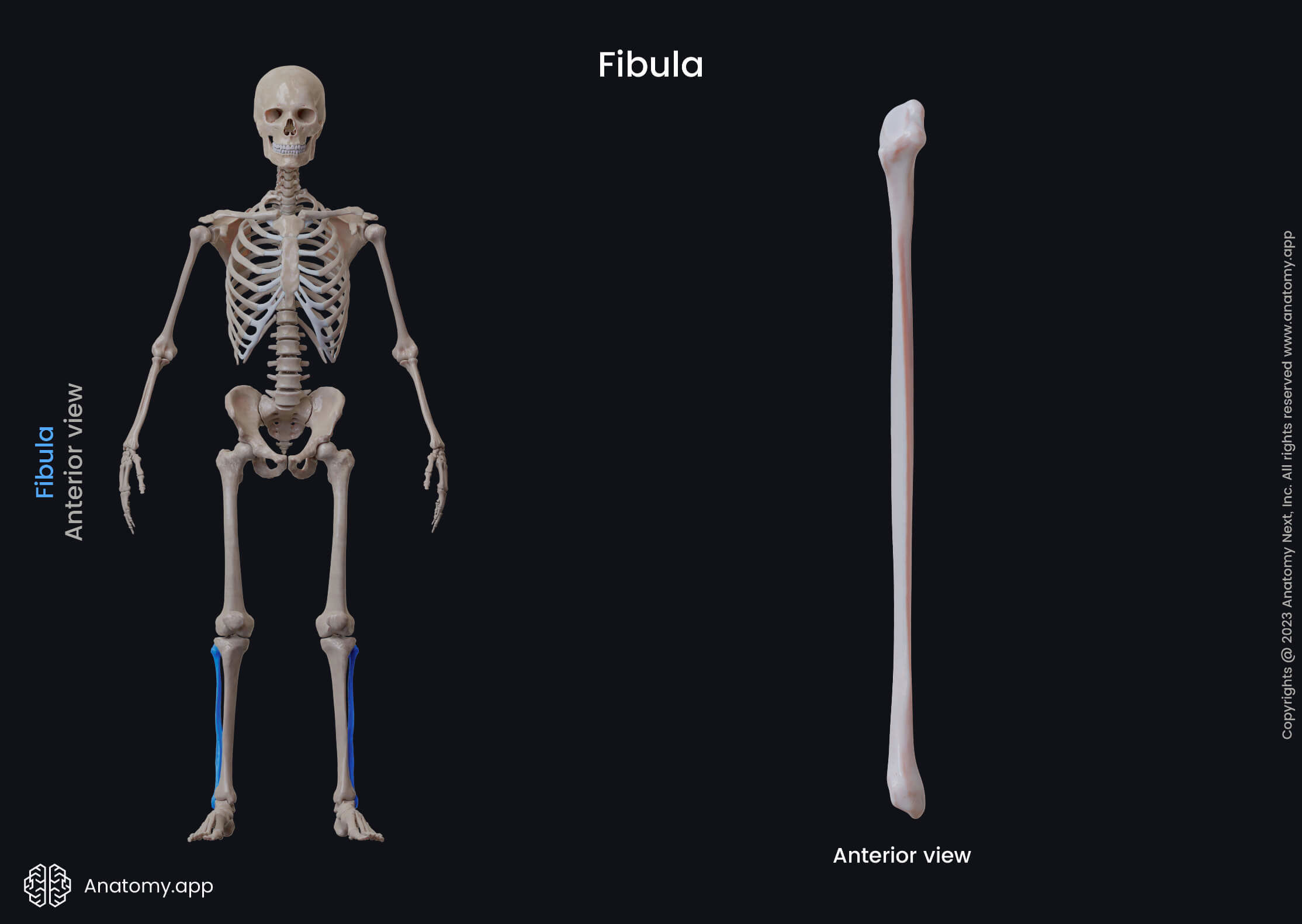
The fibula is a long bone, and as with every long bone, it also has three parts. These parts are known as follows - a diaphysis or shaft and two ends or epiphyses named the proximal and distal ends.
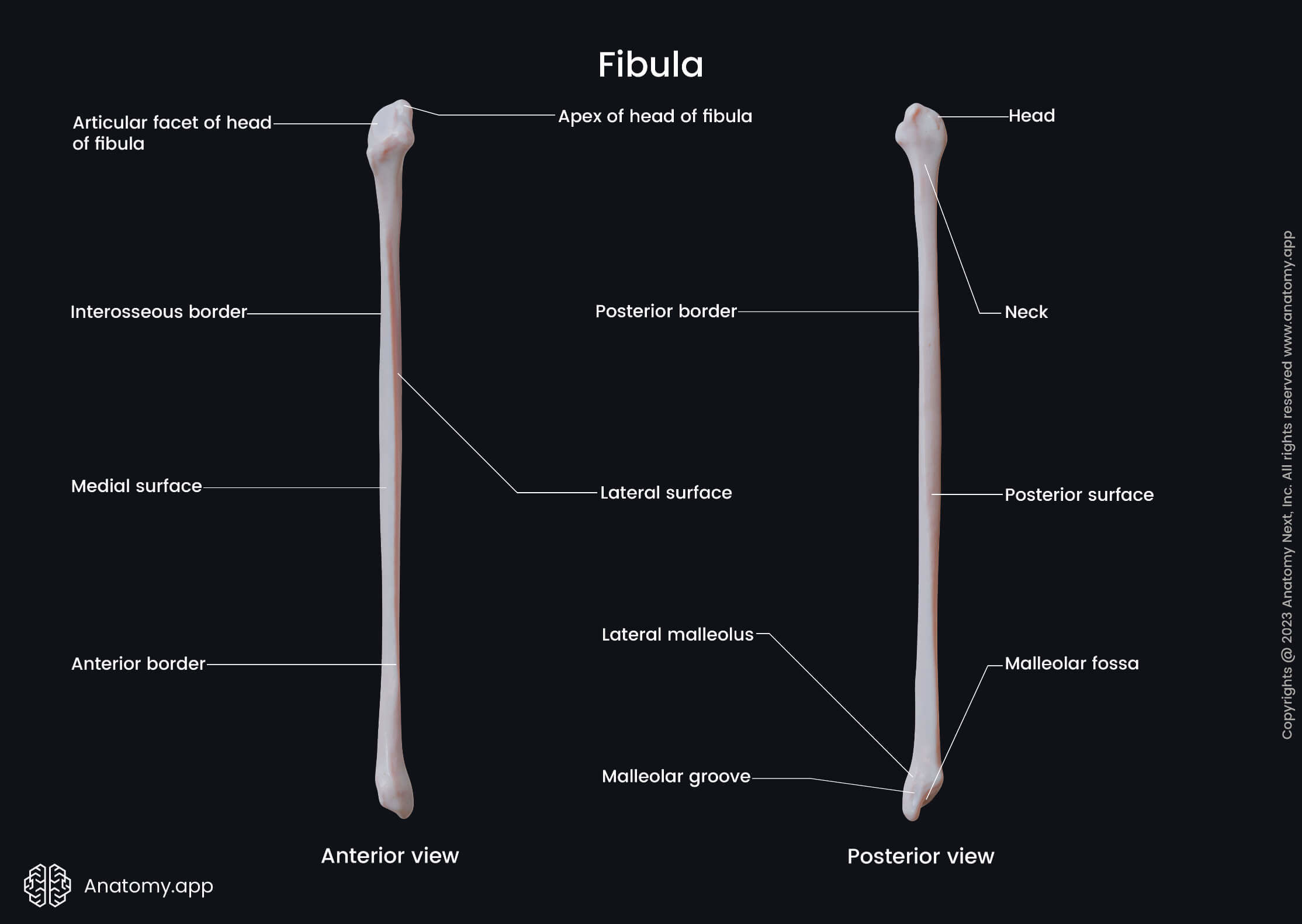
Proximal end of fibula
The proximal end or epiphysis is the upper end of the fibula that is located closer to the body. It presents the head of the fibula that features an articular surface.
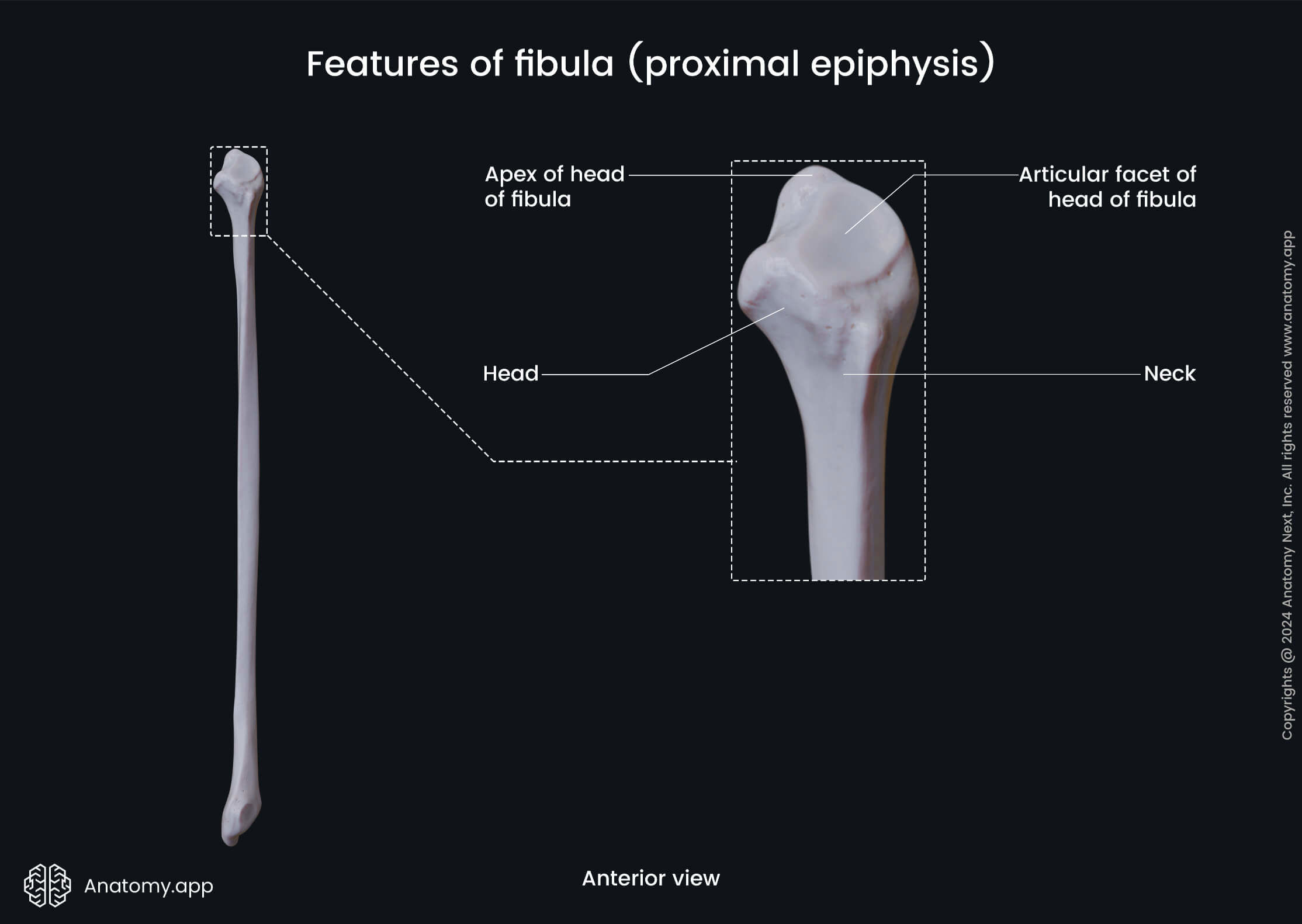
The articular surface is facing the tibia at the proximal ends of both bones. This surface articulates with the articular surface of the tibia, forming the proximal tibiofibular joint.
Shaft of fibula
The shaft or diaphysis of the fibula is the middle part of the bone. It has three surfaces - lateral, medial and posterior surfaces - and three borders - anterior, posterior and interosseous borders. The posterior surface features a ridge called the medial crest.
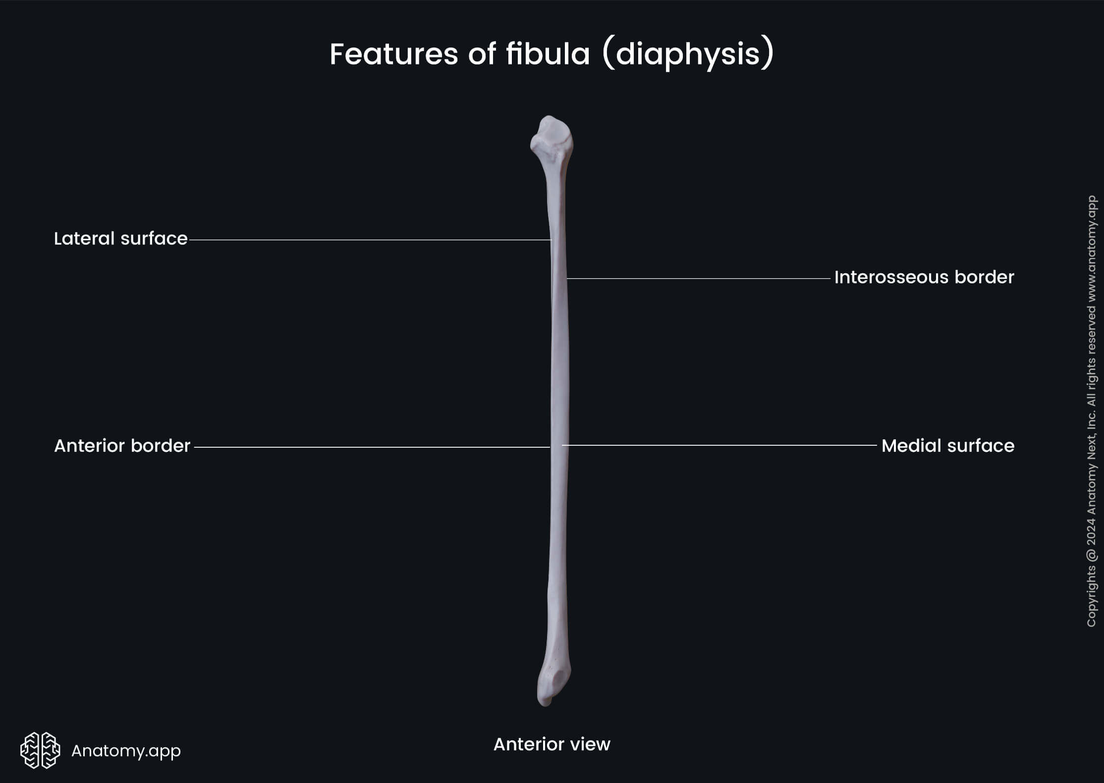
The medial crest projects on the posterior surface of the fibula at the border between the origin sites of the tibialis posterior, flexor hallucis longus and soleus muscles. It ends and continues as the interosseous border.
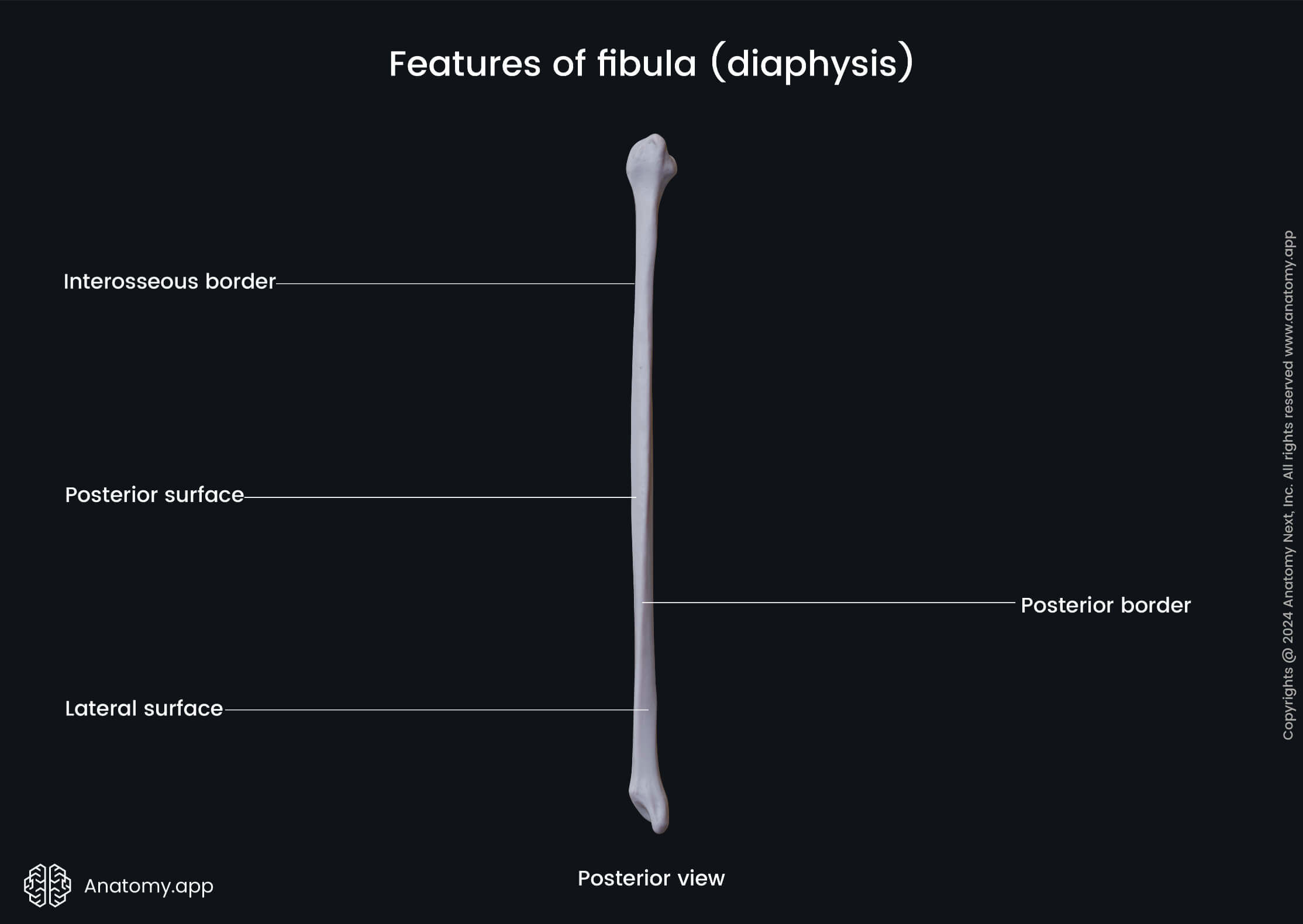
The interosseous border is the sharp medial border located between the anterior border and the medial crest. A part of the interosseous membrane located in the space between the fibula and tibia is attached to this border.
Distal end of fibula
The distal end or epiphysis of the fibula is the lower end of the bone that forms articulations with the bones of the foot. It features the lateral malleolus with an articular surface and the lateral malleolar fossa.
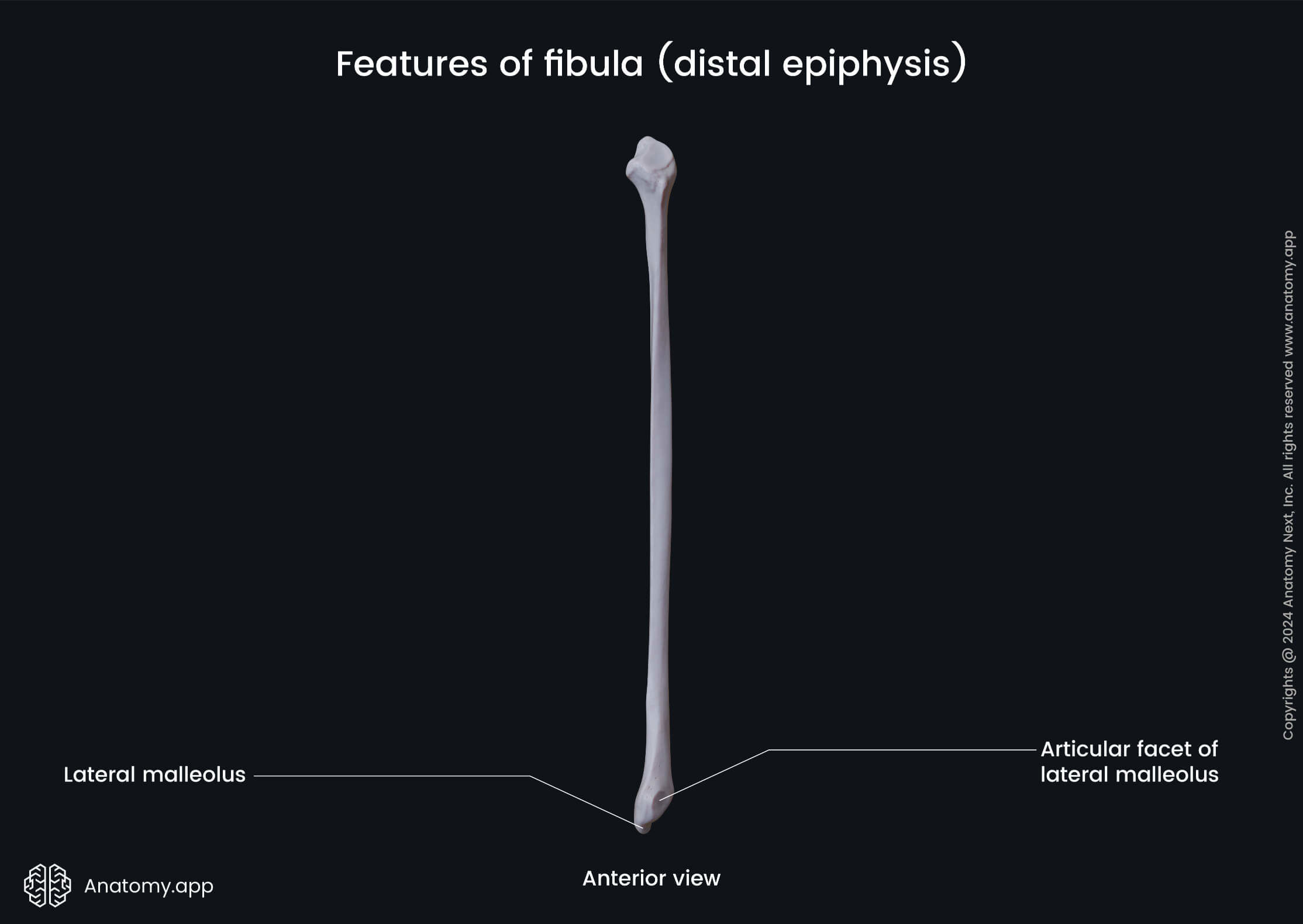
The lateral malleolus is a bony prominence on the fibular side of the ankle. The malleolar articular surface is an articular surface of the malleolus facing the talus. The articulation between both bones participates in the formation of the ankle joint.
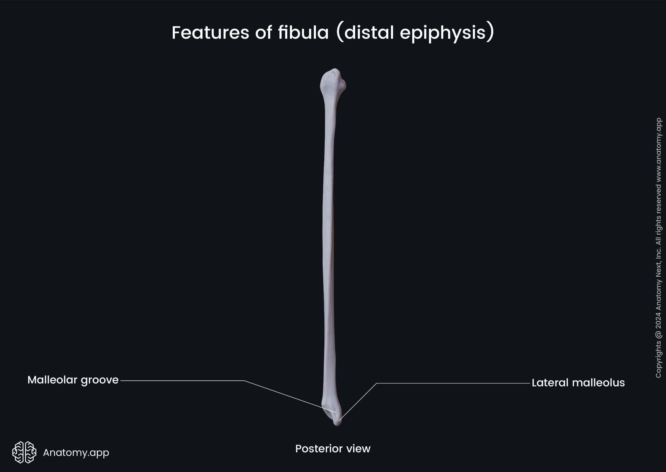
The lateral malleolar fossa is a large and rough depression on the posteromedial aspect of the lateral malleolus posterior to the articular surface. It serves as an attachment site for the posterior talofibular ligament.