- Anatomical terminology
- Skeletal system
- Skeleton of trunk
- Skull
-
Skeleton of upper limb
- Bones of shoulder girdle
- Humerus
- Bones of forearm
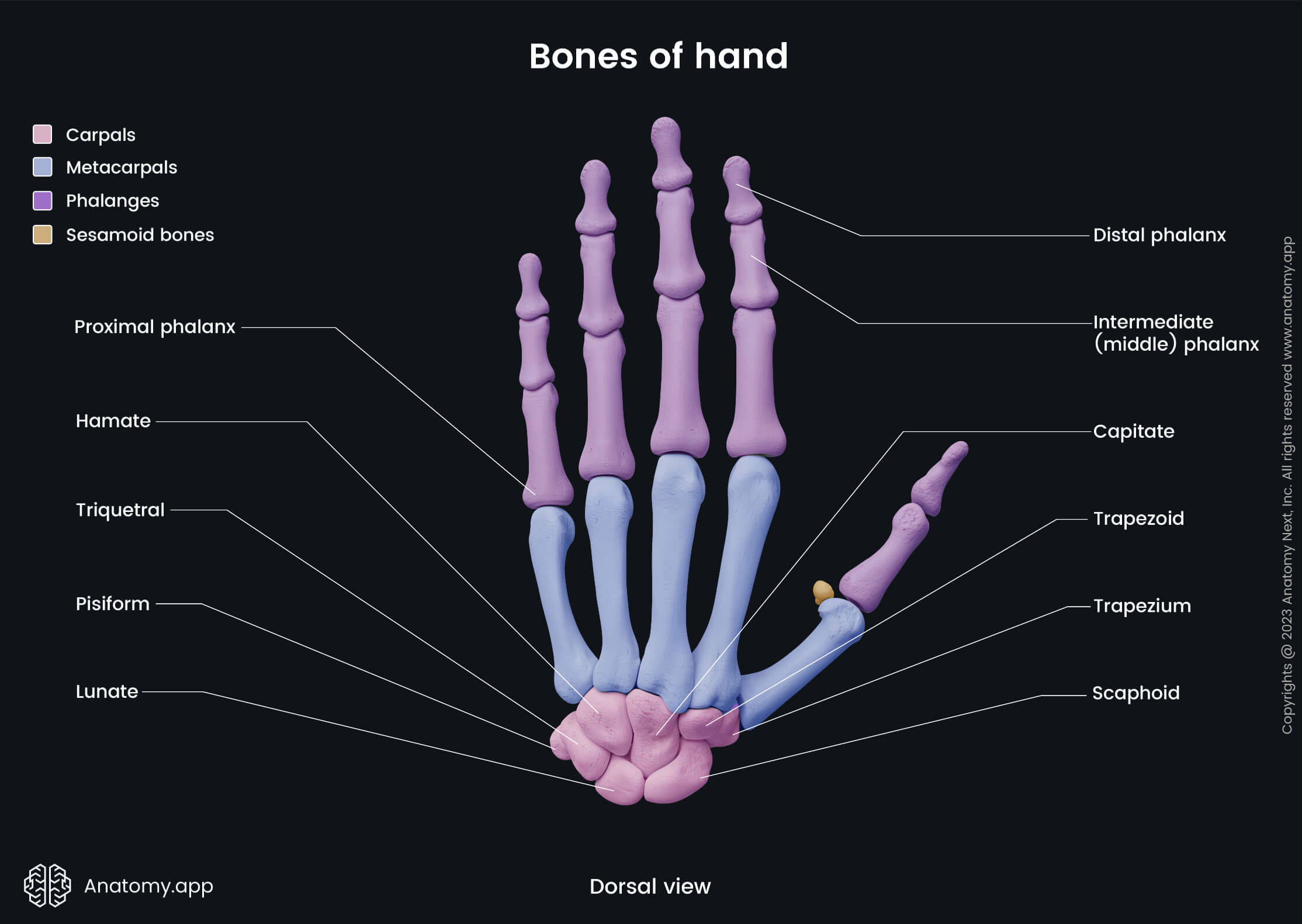
- Bones of hand
- Skeleton of lower limb
- Joints
- Muscles
- Heart
- Blood vessels
- Lymphatic system
- Nervous system
- Respiratory system
- Digestive system
- Urinary system
- Female reproductive system
- Male reproductive system
- Endocrine glands
- Eye
- Ear
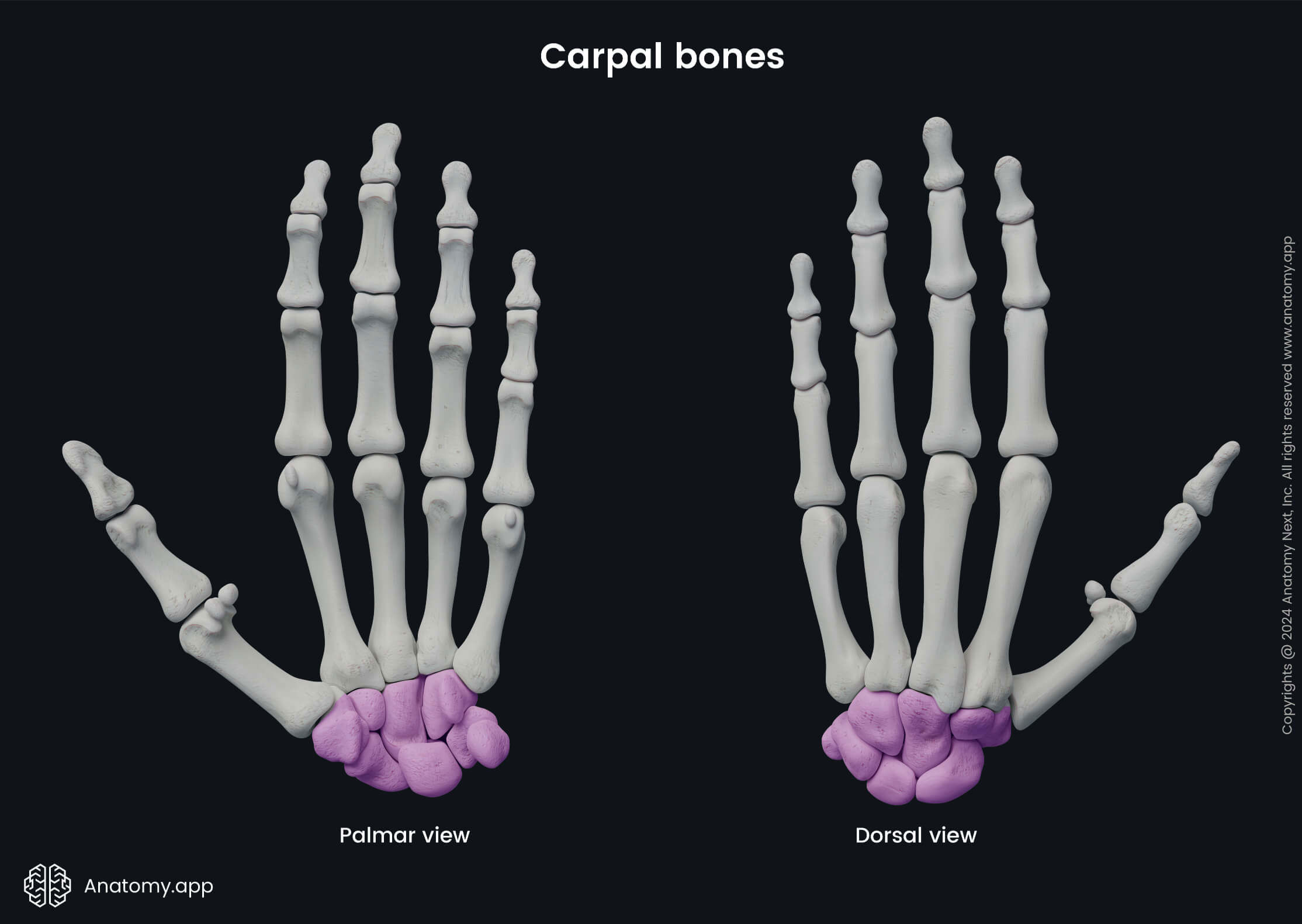
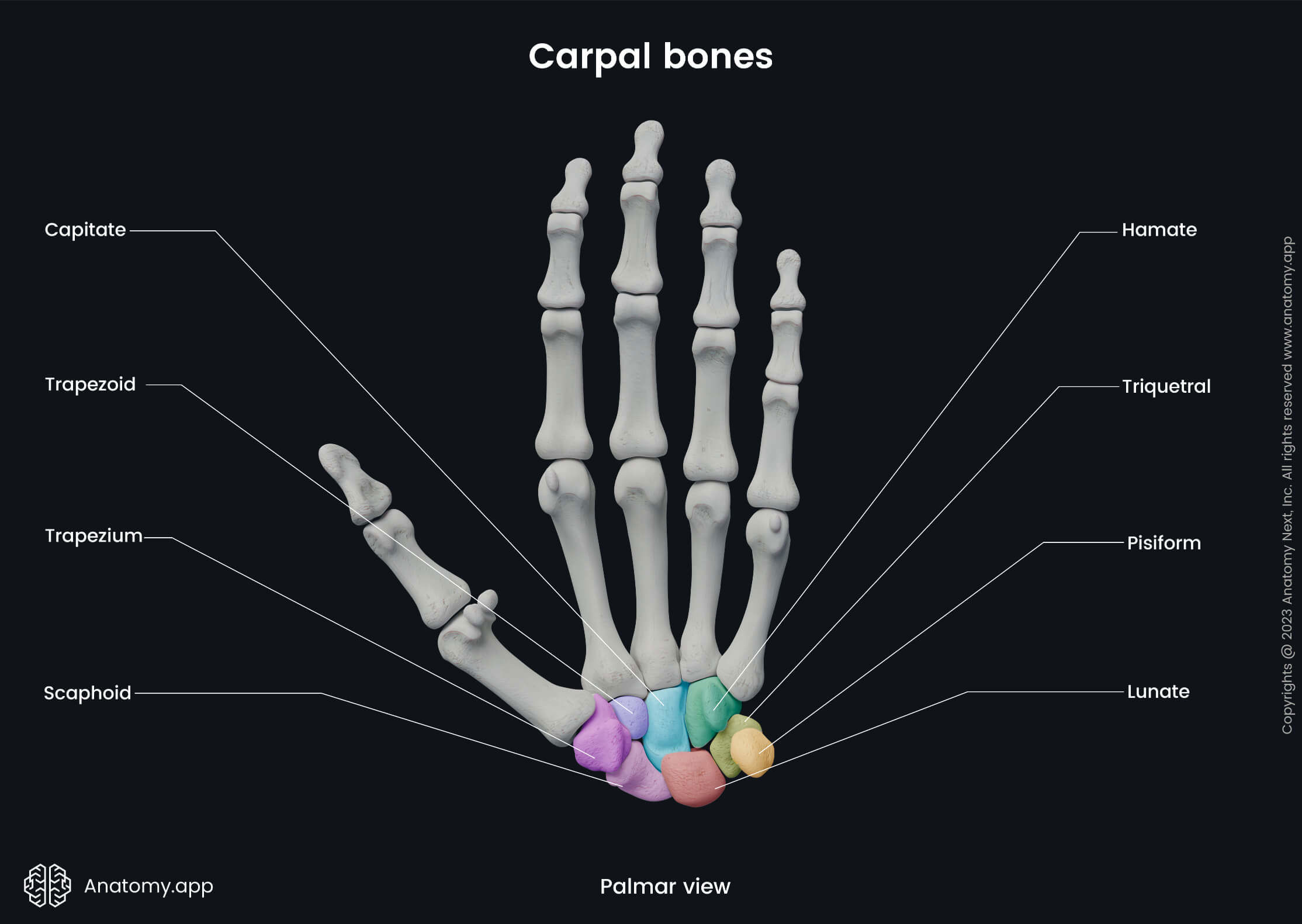
Carpal bones
The carpal bones (Latin: ossa carpi) are eight small and irregularly shaped bones located in the wrist area. These bones are arranged into two rows named the proximal and distal rows. Each row is composed of four carpal bones. The proximal row articulates with the bones of the forearm (radius and ulna), while the distal row articulates with the bases of five metacarpal bones.
The proximal row include the following carpal bones:
Four bones forming the distal row are as follows:
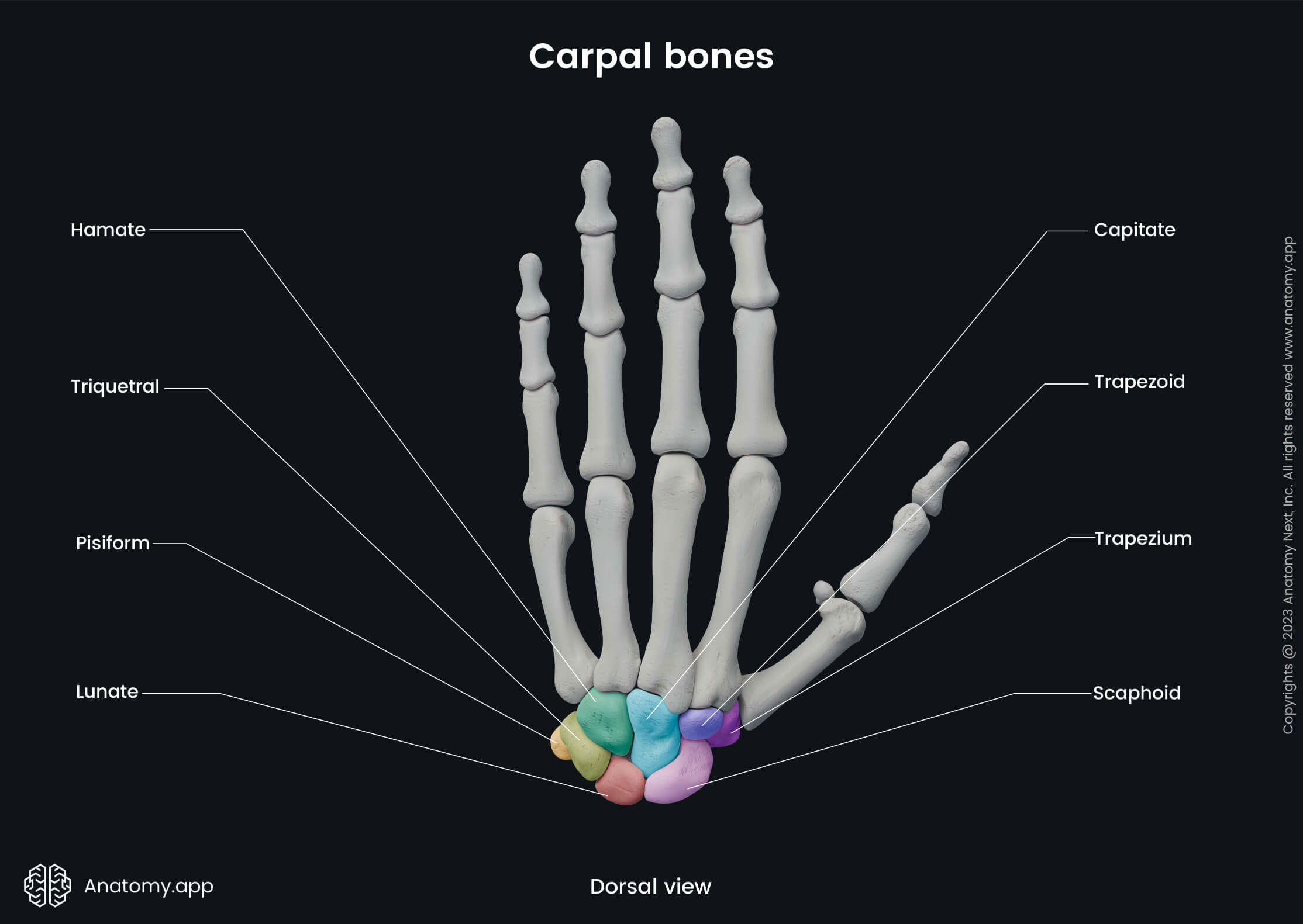
In the proximal direction, the scaphoid and lunate bones articulate with the distal end of the radius. This articulation between mentioned bones forms the wrist joint, also known as the radiocarpal joint. Distal aspects of all the carpal bones in the distal row articulate with the bases of the metacarpal bones. Overall, each carpal bone has its unique shape and multiple articular surfaces for articulation with other bones located next to them.
All carpal bones together form a transverse arch. This arch is concave on the palmar aspect, but it seems more convex proximally and concave distally. A membranous band, known as the flexor retinaculum, extends between the medial and lateral edges of the arch. Medially it attaches to the pisiform bone and the hook of hamate bone, while laterally it is attached to the trapezium bone. The space formed below the retinaculum is known as the carpal tunnel.
Scaphoid bone
The scaphoid bone (Latin: os scaphoideum) is the largest of the carpal bones located in the proximal row. It lies beneath the anatomical snuff box. The scaphoid bone presents with a tubercle, which lies subcutaneously and is easily palpable.
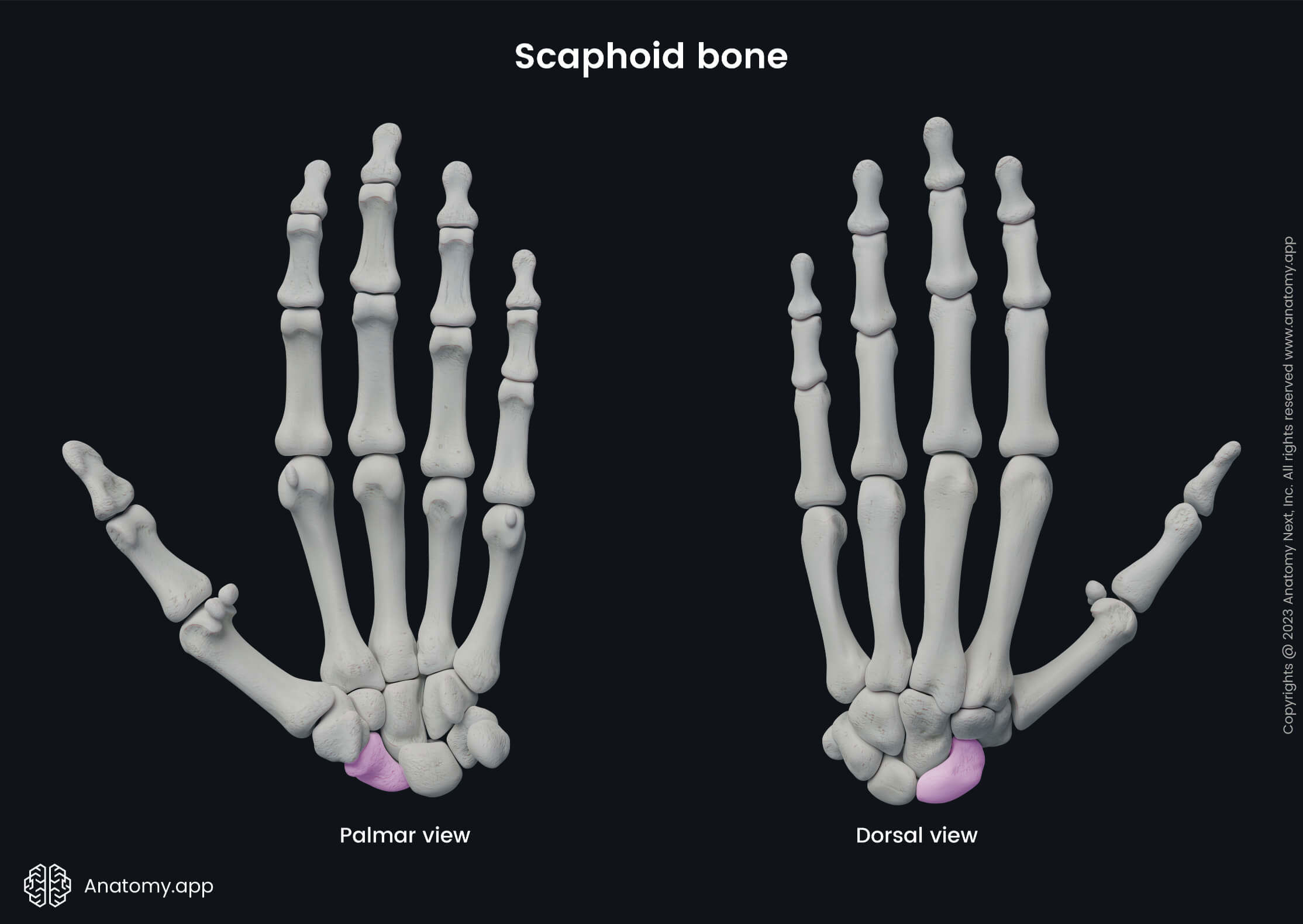
Five bones surround the scaphoid bone on the palmar aspect of the hand:
- Proximally - it is surrounded by the radius;
- Distally and laterally - next to it is the trapezium bone;
- Distally and medially - surrounded by the trapezoid bone;
- Superomedially - it connects with the lunate bone;
- Inferomedially - surrounded by the capitate bone.
Lunate bone
The lunate bone (Latin: os lunatum) is a crescent-shaped bone with a large proximal articular surface. It articulates with the radius and articular disk at the wrist joint. Sometimes the lunate bone also articulates with the hamate bone at its inferomedial angle.
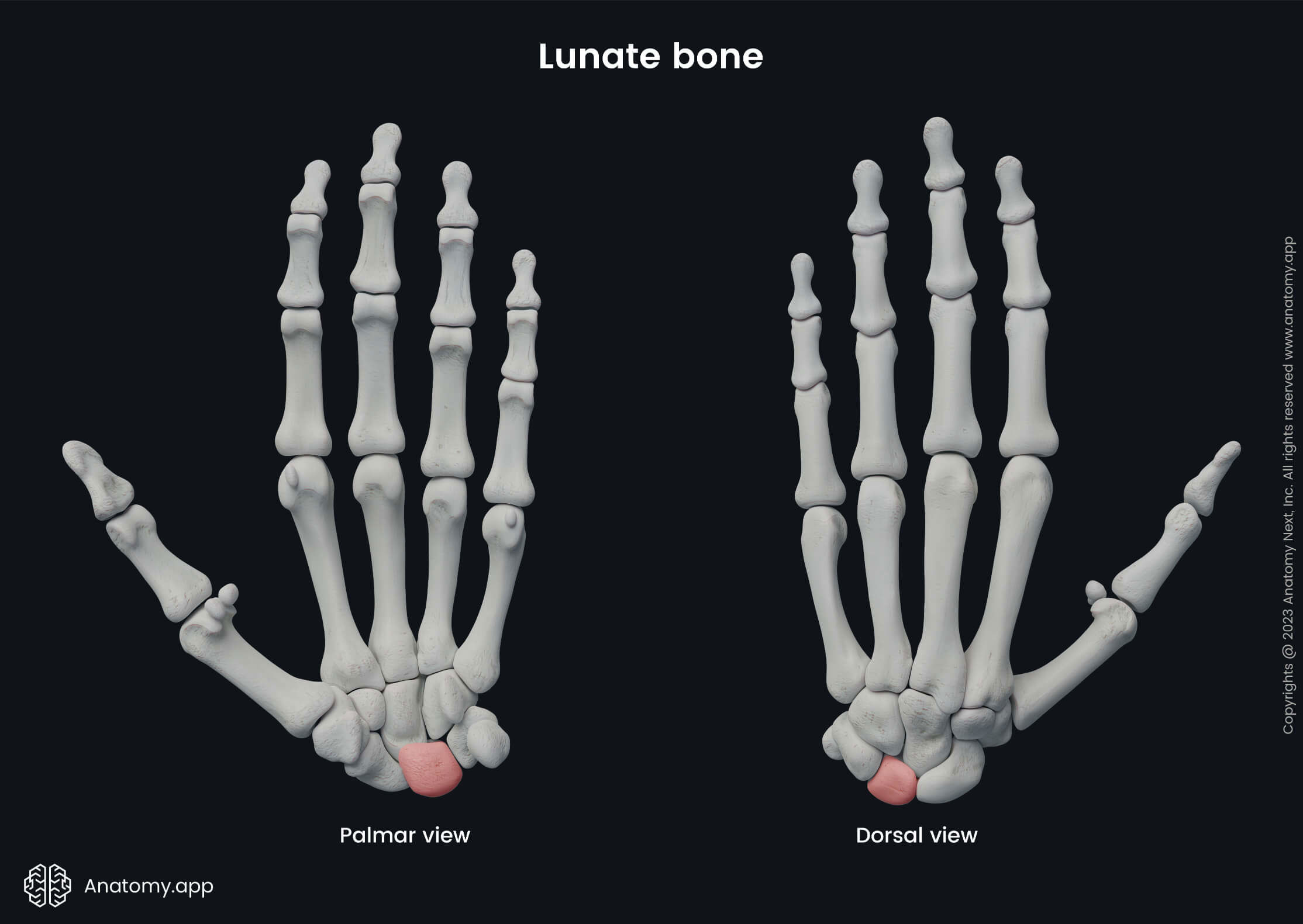
The lunate bone is connected to the following bones:
- Proximally - it is surrounded by the radius;
- Medially - next to it is the triquetral bone;
- Laterally - it connects with the scaphoid bone;
- Inferiorly - surrounded by the capitate bone.
Next to this bone is located the triquetral bone. Proximal surfaces of both bones create an articular surface that articulates with the distal end of the radius, forming the wrist joint. Besides the lunate and triquetral bones, also the scaphoid bone participates in the formation of the wrist joint.
Triquetral bone
The triquetral bone (Latin: os triquetrum, os pyramidale) is also known as triquetrum and pyramidal bone. It is a pyramidal-shaped carpal bone. From a palmar view, its apex is directed distally and medially, while its base is faced laterally.
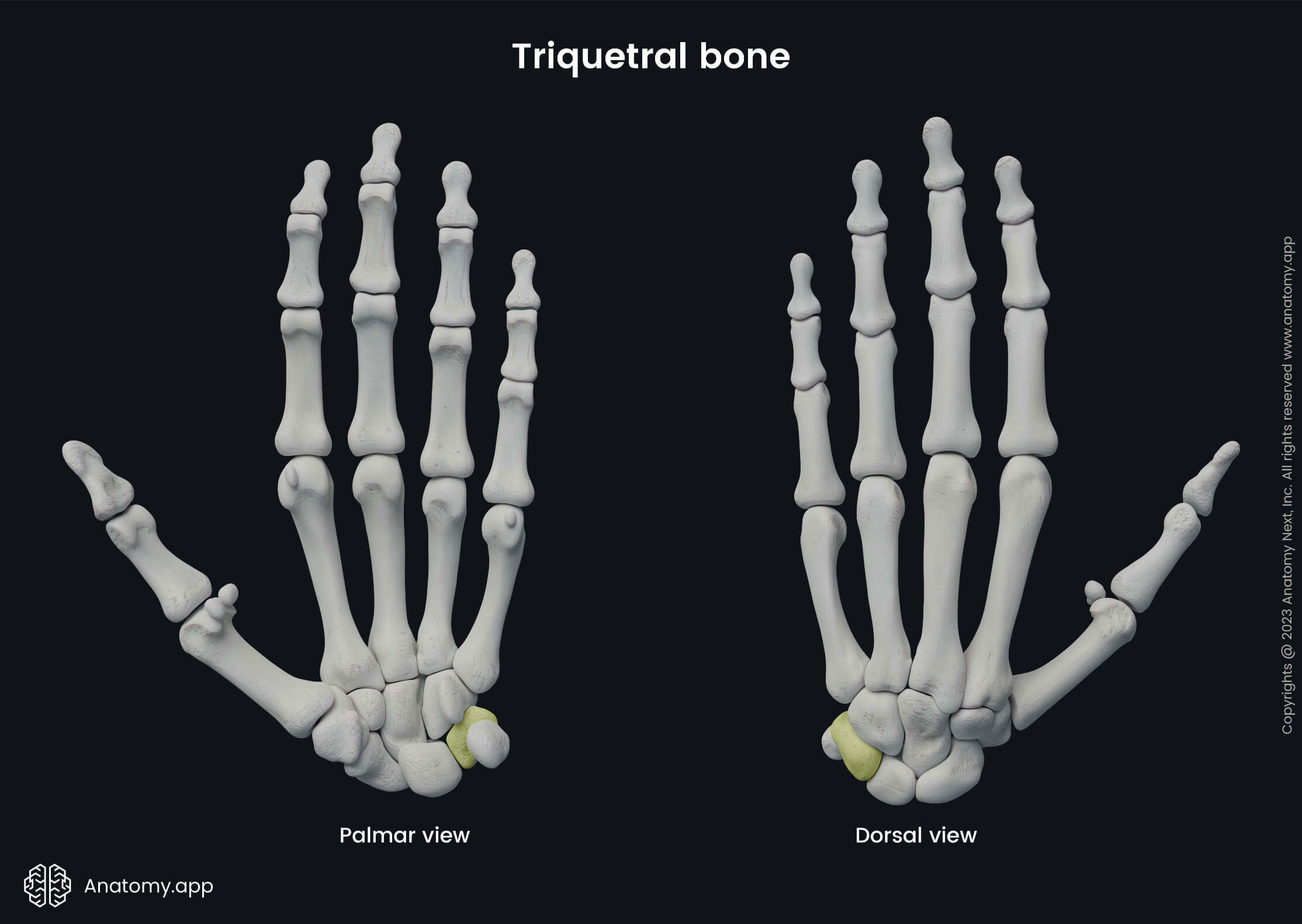
The triquetral bone is connected to the following bones:
- Distally and medially - next to it is the pisiform bone;
- Inferiorly - surrounded by the hamate bone;
- Laterally - it connects with the lunate bone.
Pisiform bone
The pisiform bone (Latin: os pisiforme) is a small and permanent sesamoid bone. It is the most medially located carpal bone from a palmar view, and it forms the ulnar border of the carpal tunnel.
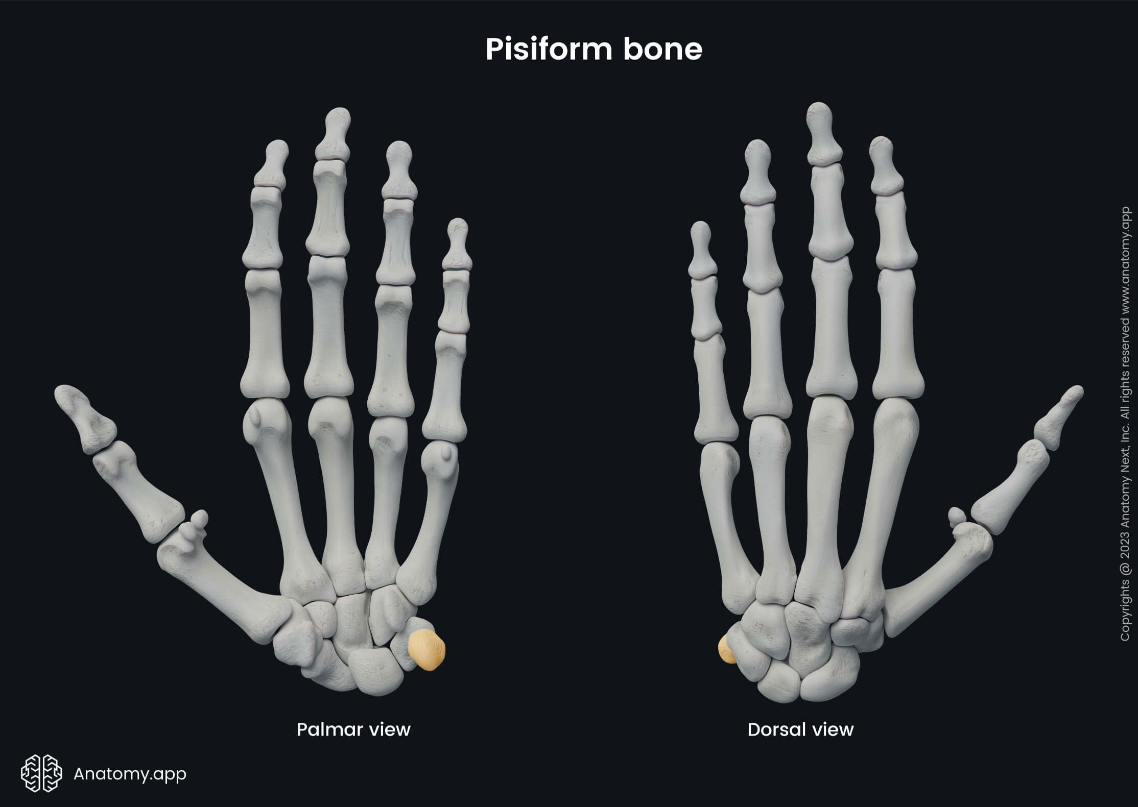
The pisiform bone lies on the triquetral bone, and the dorsal surface of the pisiform bone articulates with the ventral surface of the triquetral bone. The pisiform bone lies within the tendon of the flexor carpi ulnaris and can be palpable.
Trapezium bone
The trapezium bone or greater multangular bone (Latin: os trapezium, os multangulum majus) is the most laterally located carpal bone in the distal row when viewed from the palmar surface.
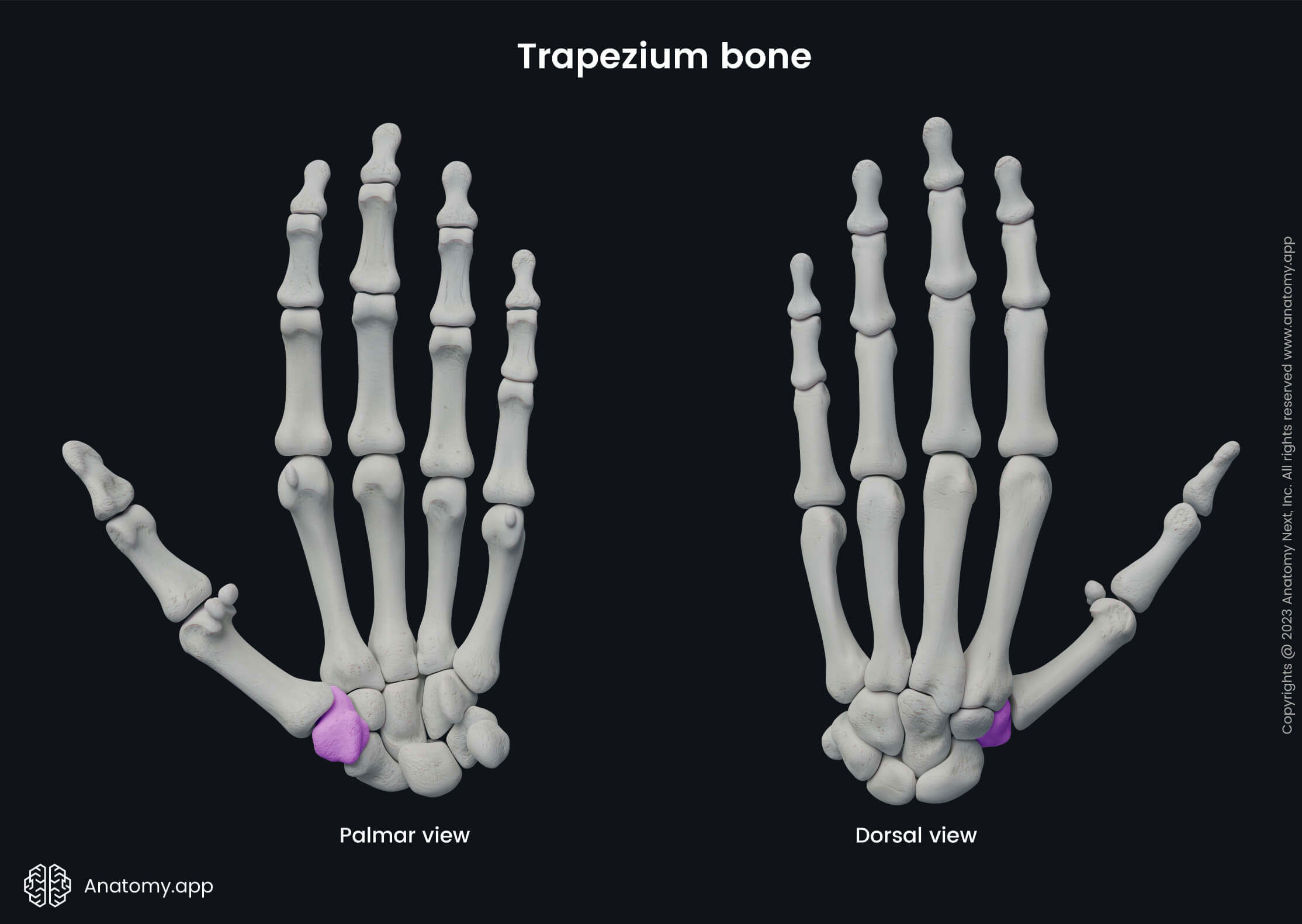
The trapezium bone has connections with several bones of the hand, including:
- Medially - next to it is the trapezoid bone;
- Superiorly - surrounded by the scaphoid bone;
- Inferolaterally - it connects with the first metacarpal bone;
- Inferomedially - it sometimes articulates with the second metacarpal bone.
The palmar surface of the trapezium bone presents with a palpable tubercle. This tubercle serves as an attachment site for the abductor pollicis brevis muscle. The medial side of the bone has a groove that gives attachment to the tendon of the flexor carpi radialis.
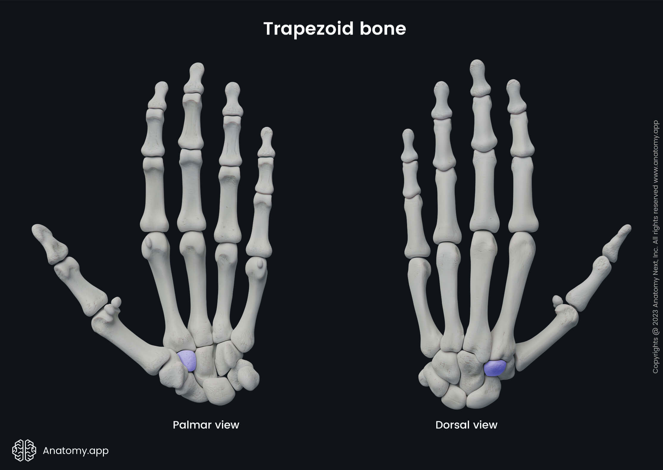
Trapezoid bone
The trapezoid bone or lesser multangular bone (Latin: os trapezoideum, os multangulum minus) is the smallest carpal bone within the distal row. It communicates with the following bones:
- Proximally - next to it is the scaphoid bone;
- Laterally - surrounded by the trapezium bone;
- Medially - it connects with the capitate bone;
- Distally - surrounded by the second metacarpal bone.
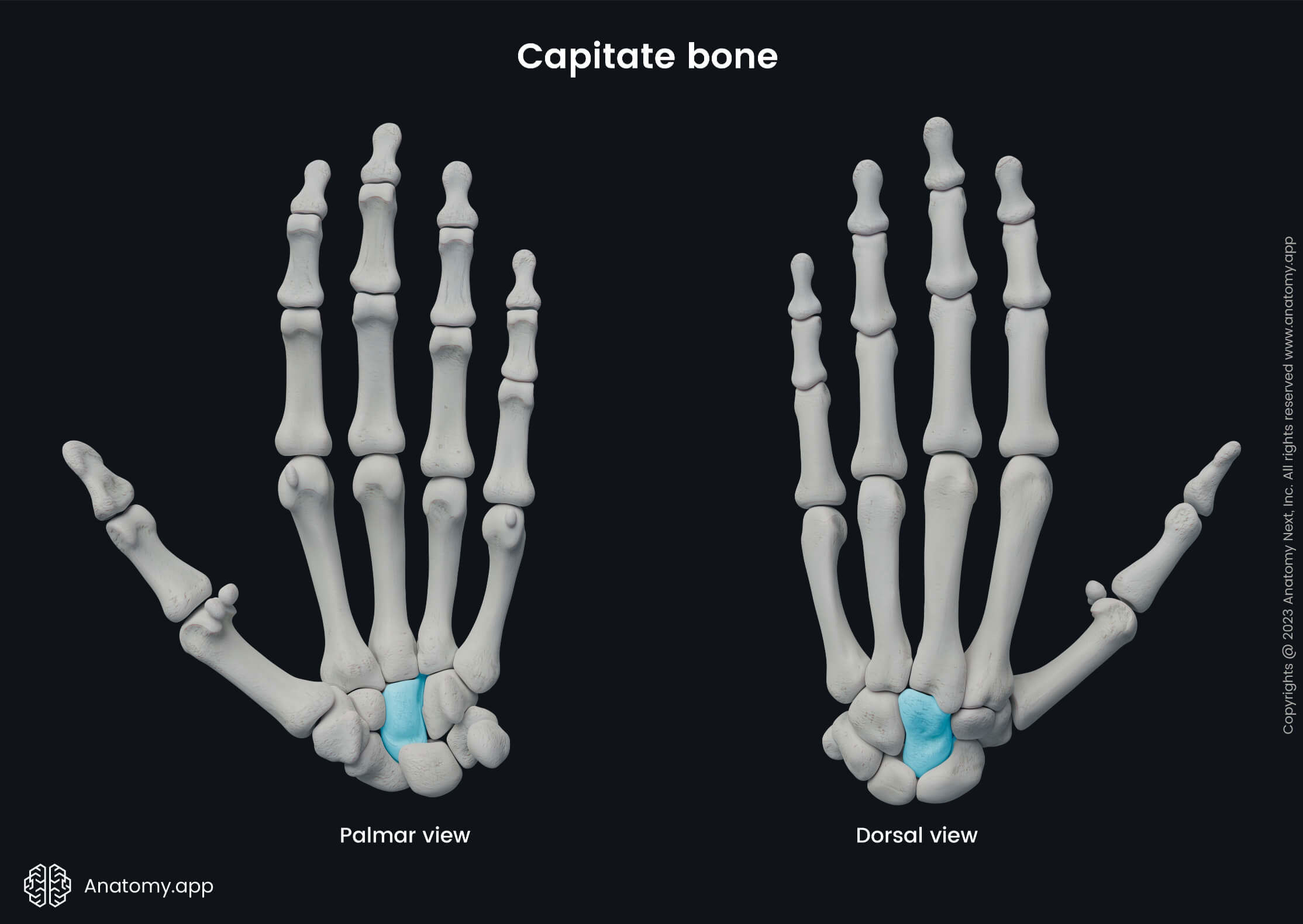
Capitate bone
The capitate bone (Latin: os capitatum) is the largest of all the carpal bones. It lies in the distal row and is surrounded by the following bones:
- Proximally - next to it is the lunate bone;
- Laterally - surrounded by the trapezoid bone;
- Medially - it connects with the hamate bone;
- Distally - surrounded by the third metacarpal bone.
Hamate bone
The hamate bone (Latin: os hamatum) is the last and most medially located carpal bone in the distal row when the hand is viewed from its palmar surface. The hamate bone lies subcutaneously and is palpable. The palmar surface of the bone contains a bony process that curves laterally, and is known as the hamulus or the hook. The flexor digiti minimi brevis and the pisohamate ligament are attached to the hamulus.
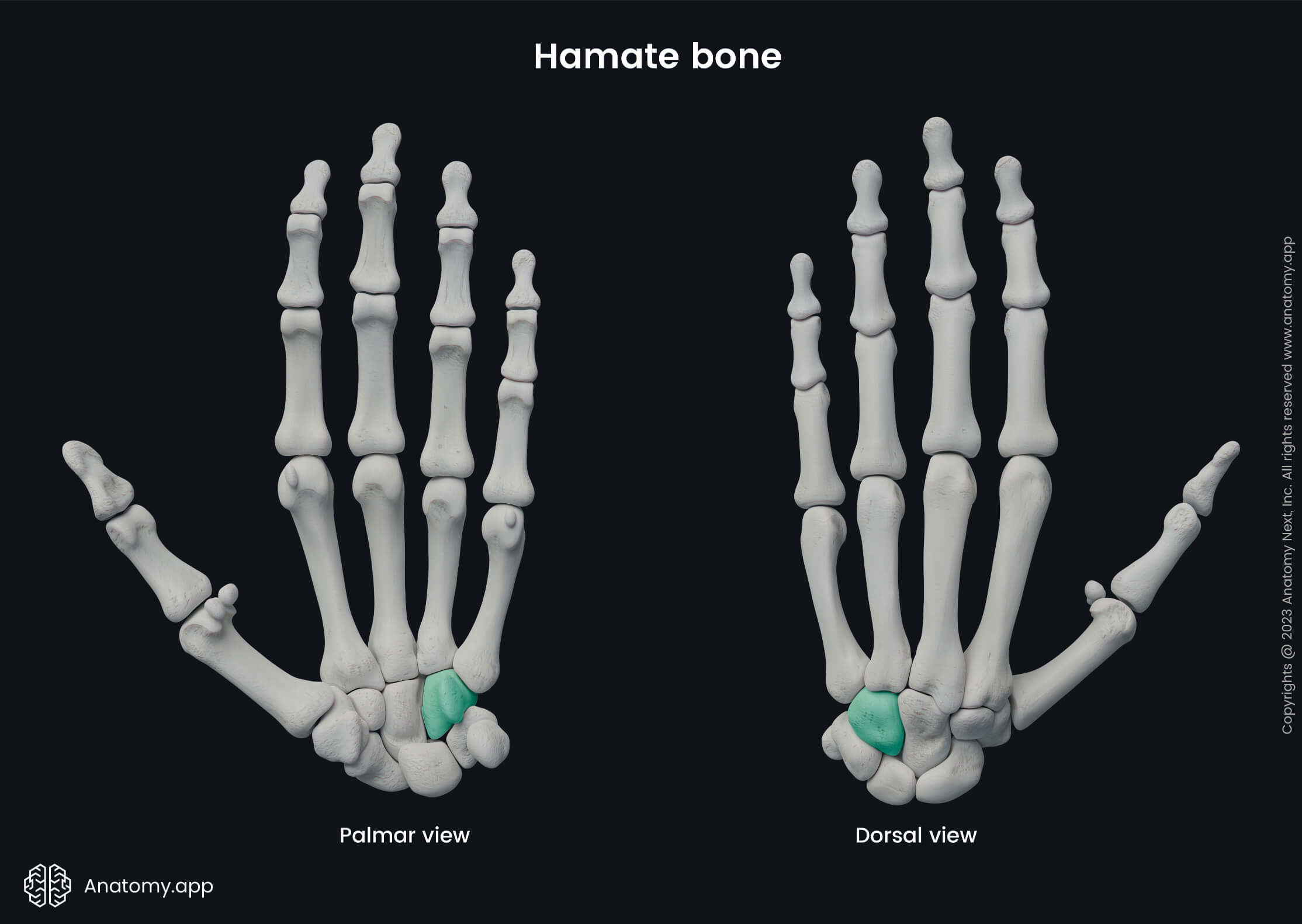
The hamate bone communicates with several bones, including:
- Proximally and laterally - surrounded by the lunate bone;
- Proximally and medially - next to it is the triquetral bone;
- Laterally - it connects with the capitate bone;
- Distally - it articulates with fourth and fifth metacarpal bones.
Anatomy.app
Contact information
- For questions regarding business inquiries. Please contact:
- info@anatomy.app