- Anatomical terminology
- Skeletal system
- Skeleton of trunk
- Skull
-
Skeleton of upper limb
- Bones of shoulder girdle
- Humerus
- Bones of forearm
- Bones of hand
- Skeleton of lower limb
- Joints
- Muscles
- Heart
- Blood vessels
- Lymphatic system
- Nervous system
- Respiratory system
- Digestive system
- Urinary system
- Female reproductive system
- Male reproductive system
- Endocrine glands
- Eye
- Ear
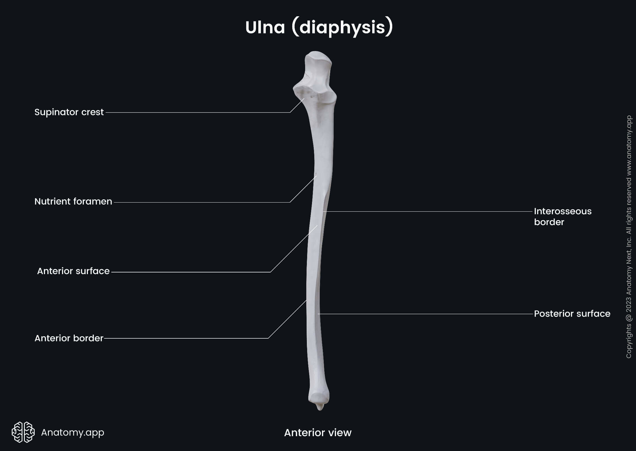
Ulna
The ulna (Latin: ulna) is one of the two long bones forming the skeleton of the forearm. It extends from the medial side of the elbow to the little finger side of the wrist. The ulna lies parallel to the radius, which is the other bone of the forearm.
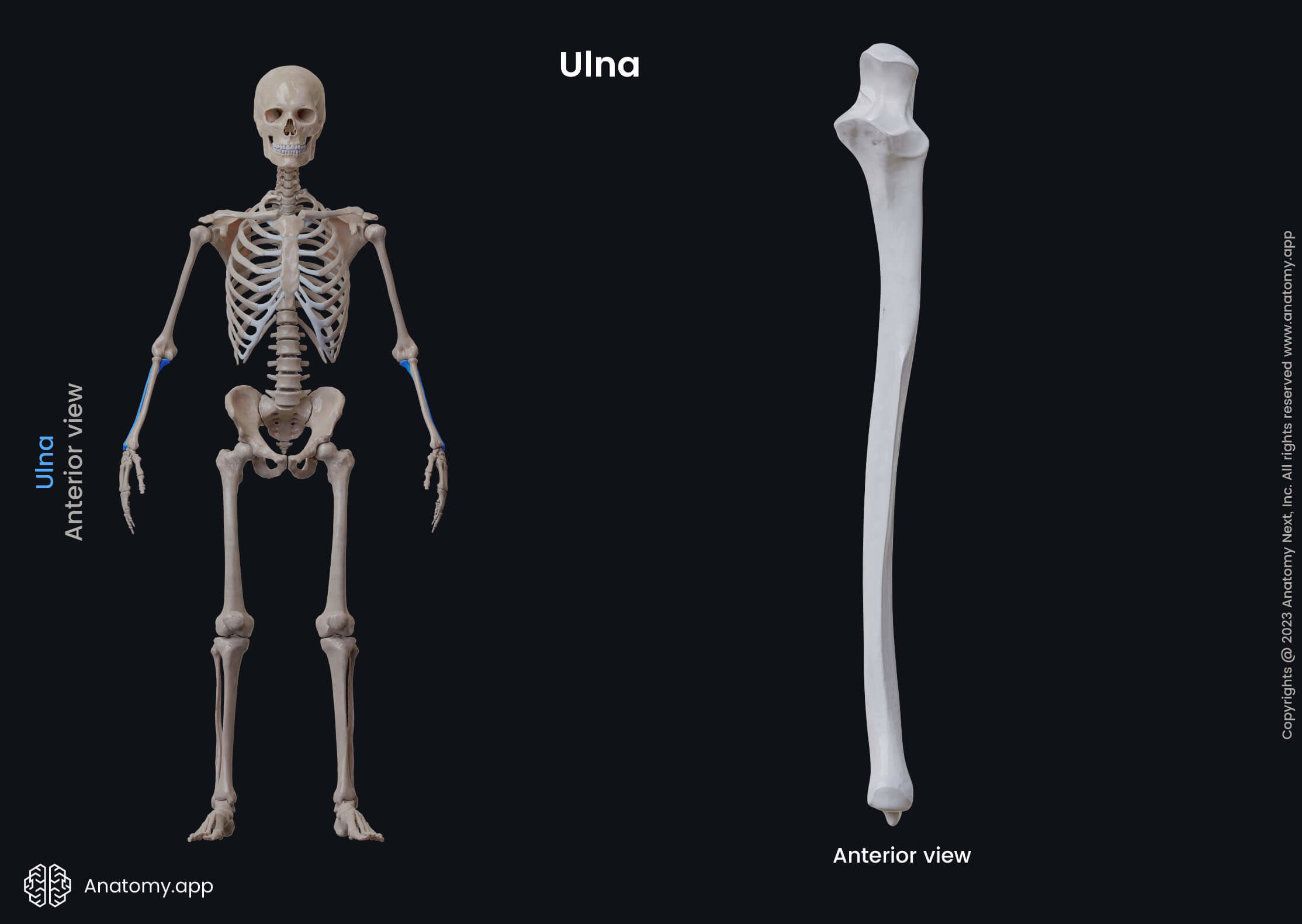
The ulna mainly acts as a stabilizing bone of the forearm. Proximally, it articulates with the humerus at the elbow joint, and it also articulates with the radius at the proximal radioulnar joint. Distally, it articulates with the radius, forming the distal radioulnar joint.
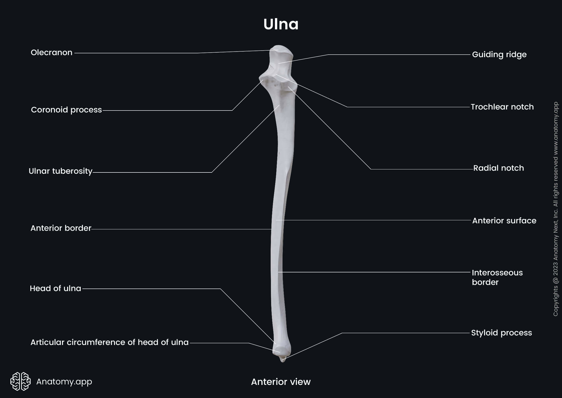
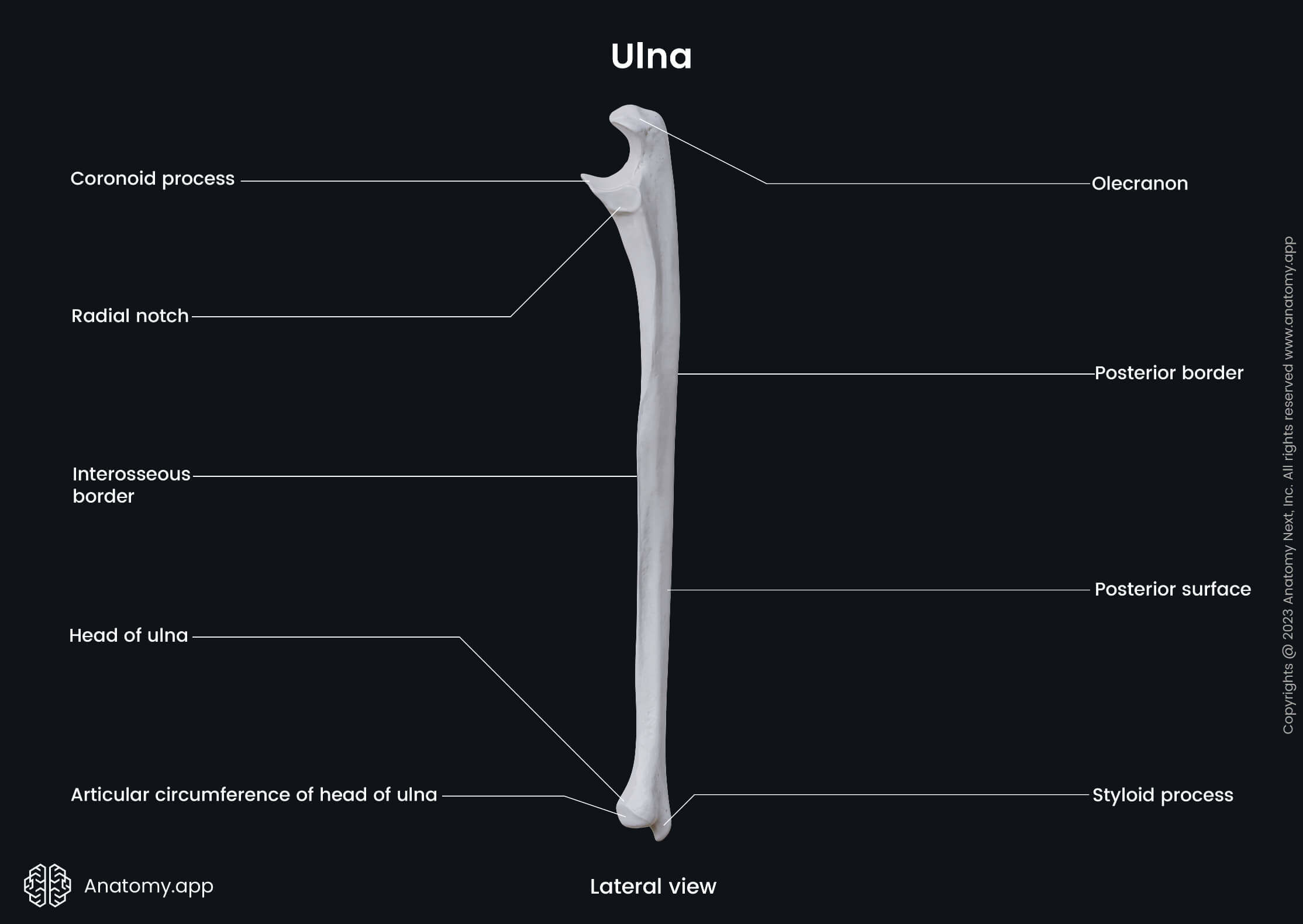
Like other long bones of the human body, the ulna also presents with three main parts: a proximal end or proximal epiphysis, a shaft or diaphysis, and a distal end or distal epiphysis. Each of these parts presents essential anatomical landmarks.
Proximal end of ulna
The proximal end or epiphysis of the ulna has two articulations. The first one participates in the formation of the elbow joint, and it is formed together with the trochlea of the humerus. The second articulation is the proximal radioulnar joint formed by the ulna and the head of the radius. The surface of the proximal ulna contains bony prominences for muscle attachment, which enable movements at the elbow joint. The proximal end contains several essential anatomical landmarks of the ulna, and these landmarks include the olecranon, coronoid process, trochlear notch, radial notch and the tuberosity of the ulna.
The olecranon is a large, thick and curved bony projection of the proximal end of the ulna that extends in a proximal direction on the posterior aspect of the bone. It also forms a part of the trochlear notch. The olecranon can be palpated as the most pointed portion of the elbow behind it. The olecranon is bent forward at the summit, thus presenting a prominent lip. The mentioned lip of the olecranon is received into the olecranon fossa of the humerus during the extension of the forearm. The superior surface of the olecranon serves as an attachment site for the triceps brachii muscle.
The coronoid process of the ulna is a triangular eminence that projects forward from the anterior proximal part of the ulna, and it forms a part of the trochlear notch. The apex of this process is received into the olecranon fossa of the humerus during the flexion of the forearm. Its medial surface serves as the attachment site for part of the ulnar collateral ligament. The medial surface is also the origin site for one head of the flexor digitorum superficialis, part of the flexor digitorum profundus, and one head of the pronator teres muscle. Also, the flexor pollicis longus muscle frequently arises from the lower part of the coronoid process.
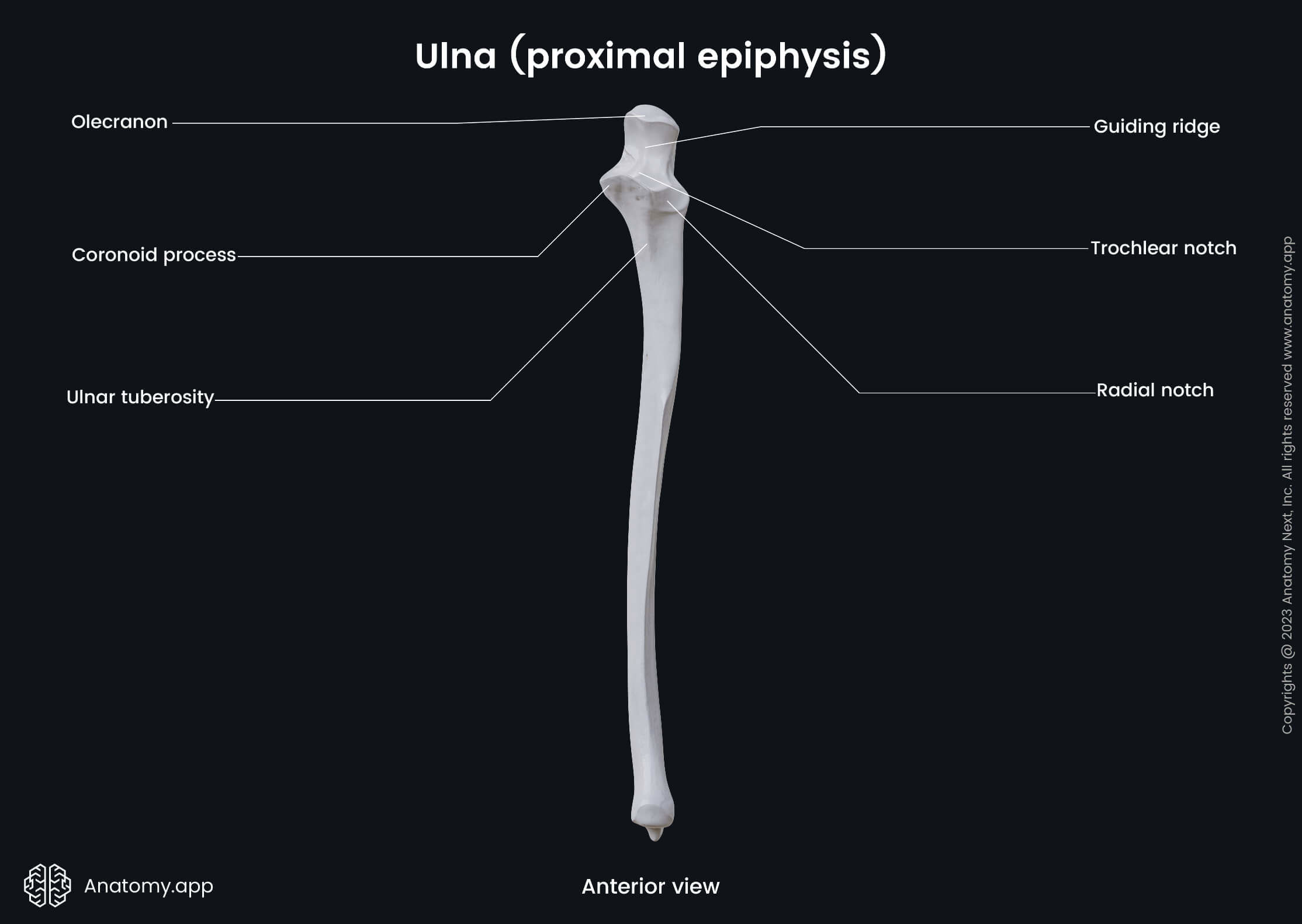
The trochlear notch or semilunar notch is a significant depression found on the proximal end and formed by the olecranon and the coronoid process. It is wrench-shaped, and in it fits the trochlea of the humerus, forming a part of the elbow joint. The radial notch of the ulna is a narrow, oblong depression located on the lateral surface of the coronoid process, laterally to the trochlear notch. It receives the articular circumference of the head of the radius at the proximal radioulnar joint. The tuberosity of the ulna is a rough eminence located immediately below the coronoid process. A part of the brachialis muscle inserts on this tuberosity.
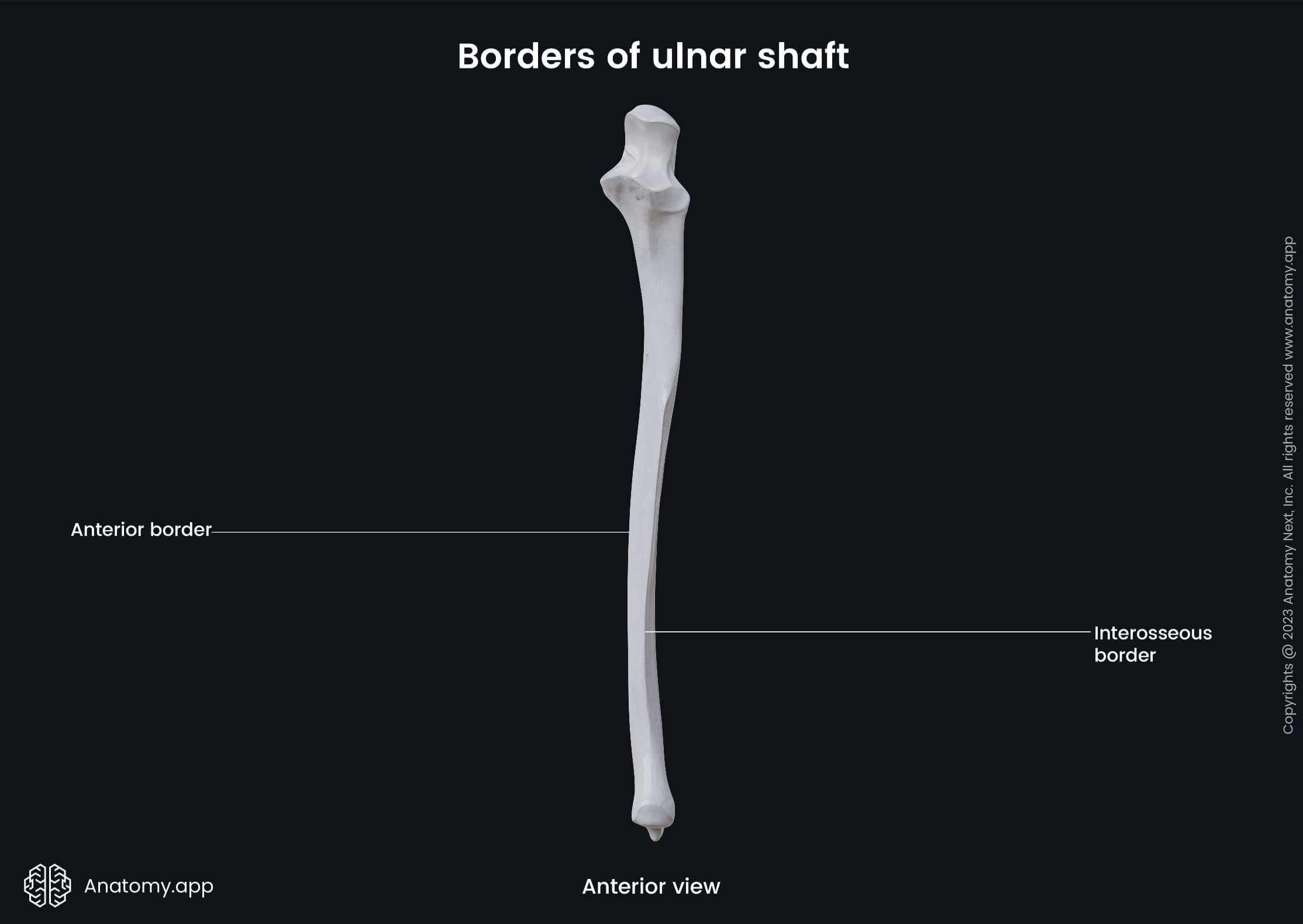
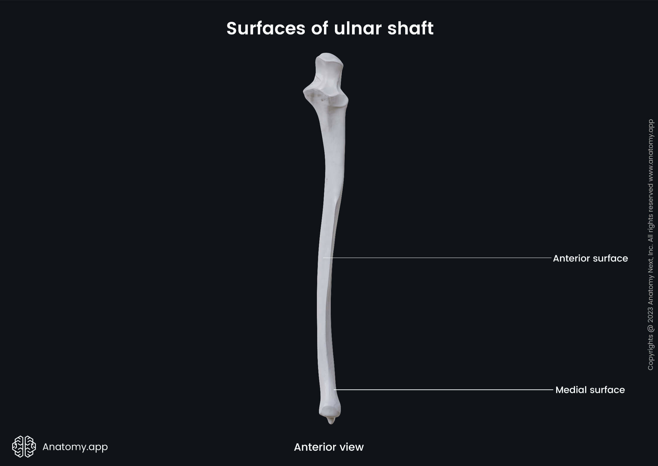
Ulnar shaft
The shaft of the ulna presents with a triangular shape when viewed in cross-section studies. It is also referred to as the ulnar shaft or diaphysis.
The ulnar shaft has three borders - anterior, posterior and interosseous borders - and three surfaces - anterior, posterior and medial surfaces. Distally, the diameter of the shaft decreases.
Borders of ulnar shaft
The anterior border is also known as the volar border. Its upper part is well defined, while its middle portion is smooth and rounded, giving origin to the flexor digitorum profundus. The lower fourth of this border gives origin to the pronator quadratus muscle.
The posterior border (also dorsal border) posteriorly is palpable along the entire length of the forearm. It serves as an attachment site for an aponeurosis that gives a common origin to the flexor carpi ulnaris, extensor carpi ulnaris and flexor digitorum profundus muscles.
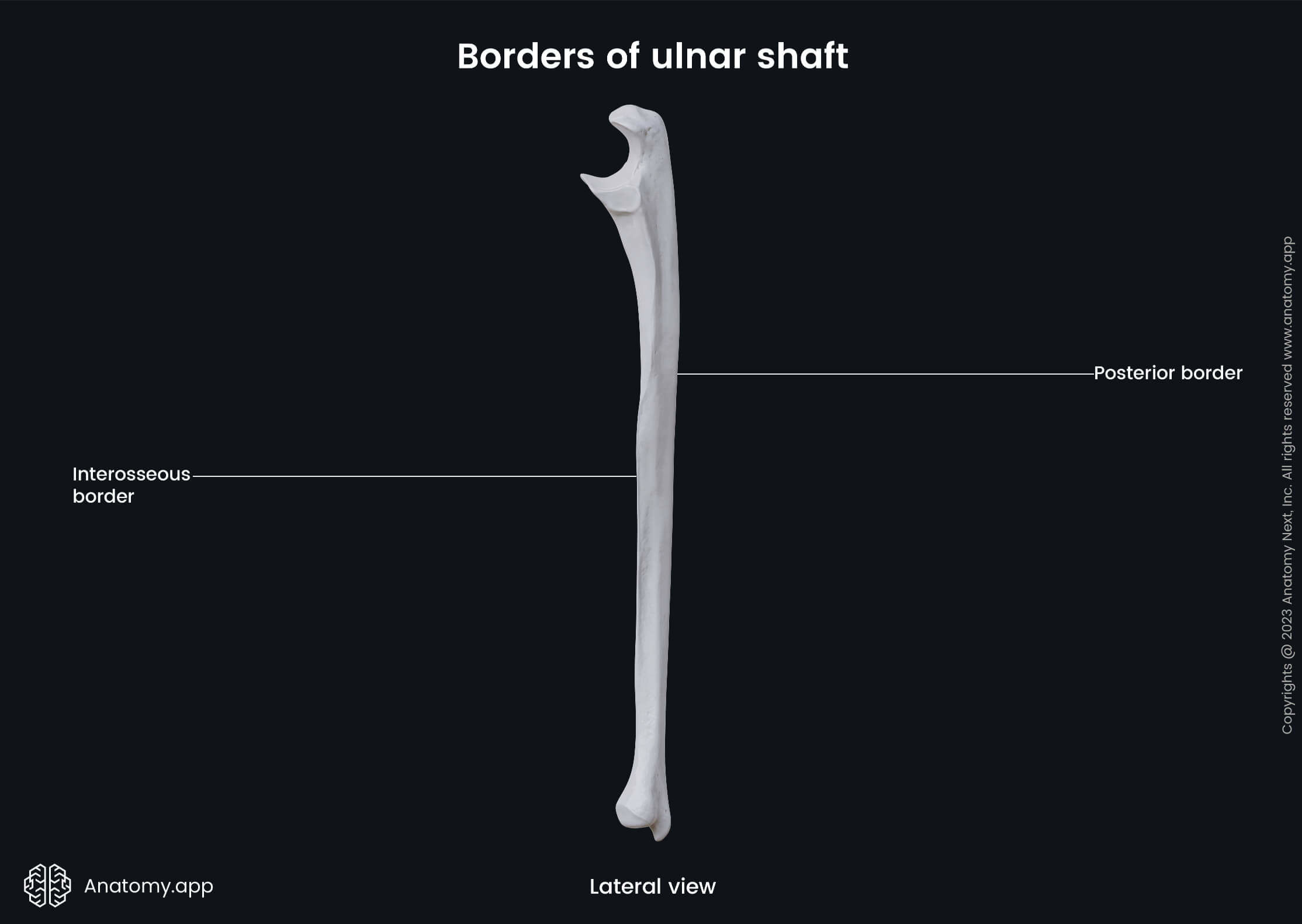
The interosseous border is also referred to as the interosseous crest. This border is sharper than others, and it is outward-facing. The interosseous border serves as the site for attachment of the interosseus membrane, extending between the two forearm bones.
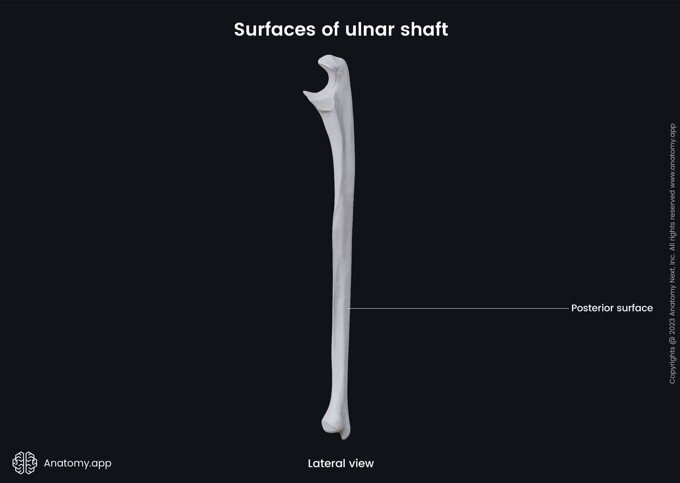
Surfaces of ulnar shaft
The anterior surface (also volar surface) serves as the origin site for the flexor digitorum profundus muscle, and distally it is covered by the pronator quadratus muscle. The flexor digitorum profundus also originates from the upper three-thirds of the medial surface.
The posterior surface of the ulnar shaft is also known as the dorsal surface. This surface is an attachment site for several muscles, including the anconeus, supinator, abductor pollicis longus, extensor pollicis longus, and extensor indicis muscles.
Distal end of ulna
The distal end or epiphysis of the ulna is a part of the ulna located closer to the wrist. It is smaller in diameter than the proximal end. The distal end of the ulna terminates with a rounded head and a distal projection known as the ulnar styloid process. The head of the ulna also contains an articular circumference for articulation with the carpal bones and radius.
The head of the ulna contains an articular surface, part of which is directed downward. It articulates with the upper surface of the triangular articular disk that separates it from the wrist joint. The remaining portion of the head is directed laterally. It is narrow, convex, and forms the distal radioulnar joint by articulating with the ulnar notch of the radius.
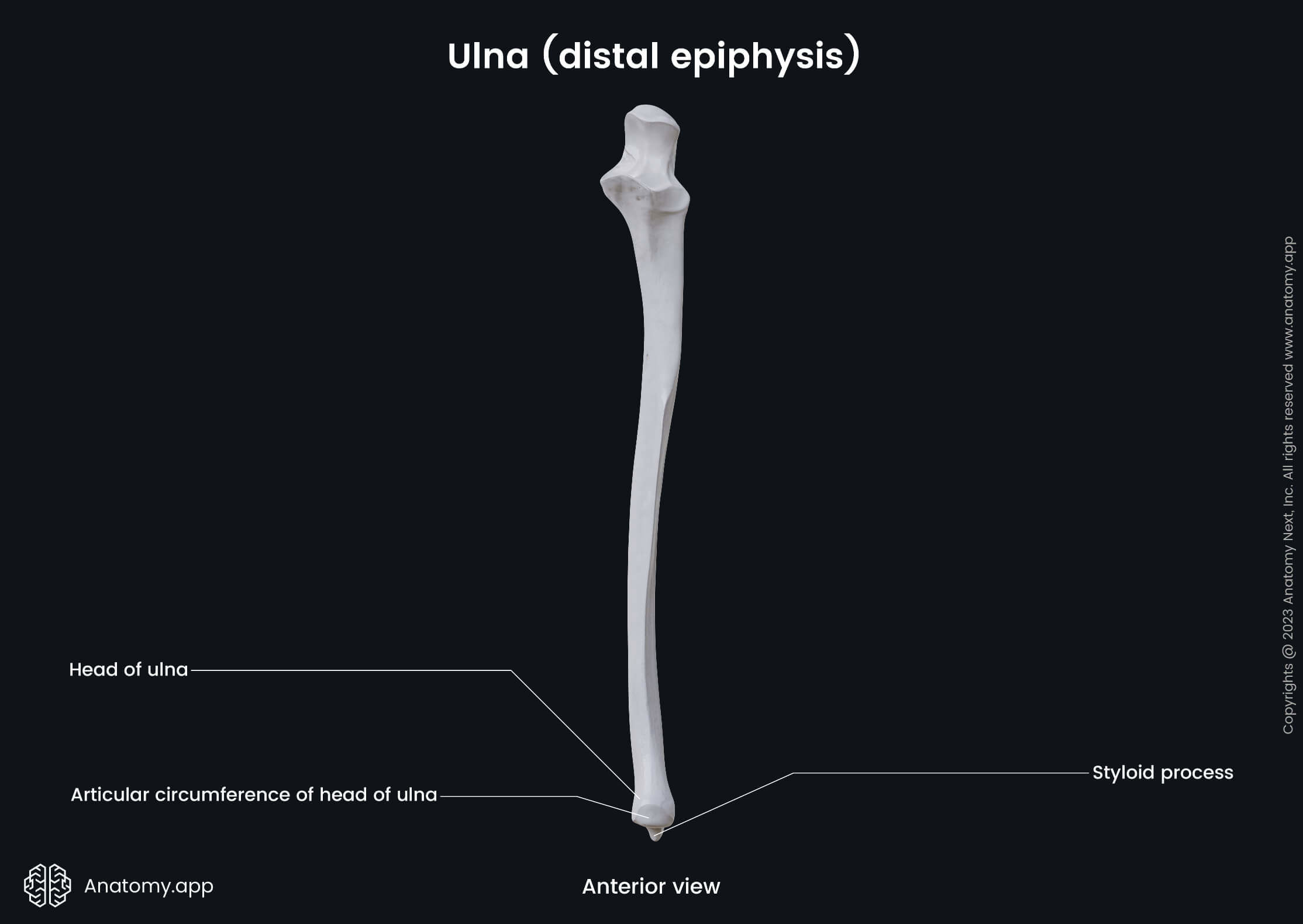
The styloid process of the ulna is a downward projection from the medial and posterior parts of the bone. The end of the styloid process is rounded, and it serves as an attachment site for the ulnar collateral ligament of the wrist joint. Just above the styloid process on the medial aspect of the ulna, to the bone is attached the pronator quadratus muscle.
Anatomy.app
Contact information
- For questions regarding business inquiries. Please contact:
- info@anatomy.app