- Anatomical terminology
- Skeletal system
- Joints
- Classification of joints
- Joints of skull
- Joints of spine
- Joints of lower limb
- Joints of pelvis
- Hip joint
- Knee joint
- Tibiofibular joints
- Joints of foot
- Muscles
- Heart
- Blood vessels
- Lymphatic system
- Nervous system
- Respiratory system
- Digestive system
- Urinary system
- Female reproductive system
- Male reproductive system
- Endocrine glands
- Eye
- Ear
Subtalar joint
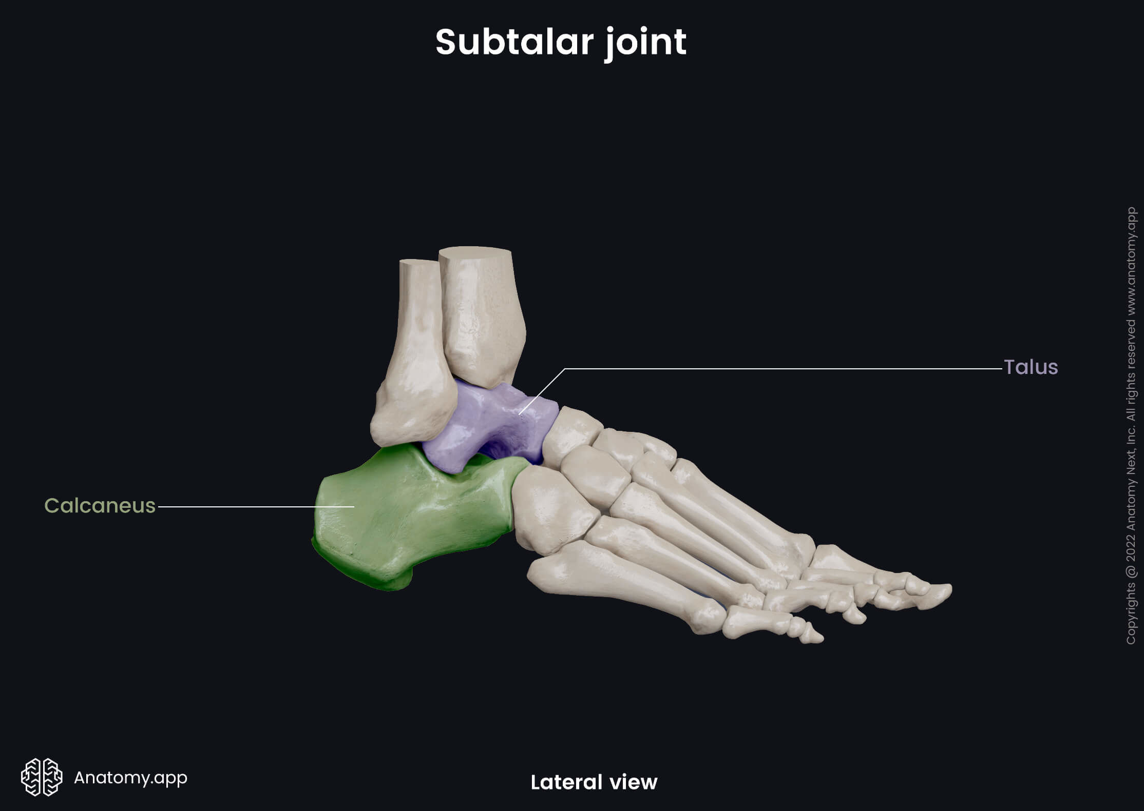
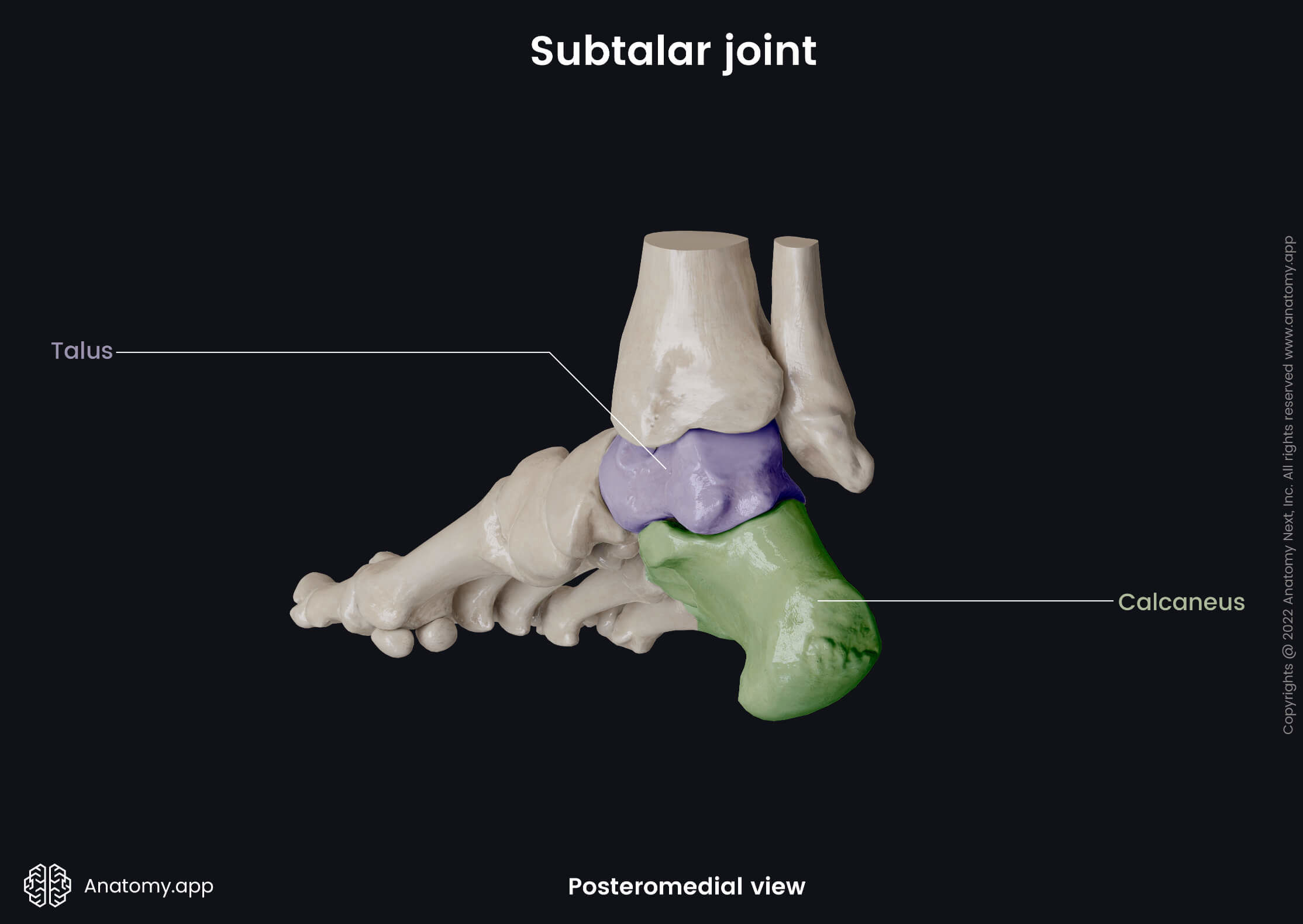
The subtalar joint (also known as the talocalcaneal joint; Latin: articulatio subtalaris) is a synovial plane type articulation located between the tarsal bones of the foot. This joint is formed between two bones of the foot - the talus and calcaneus.
Articulating structures of subtalar joint
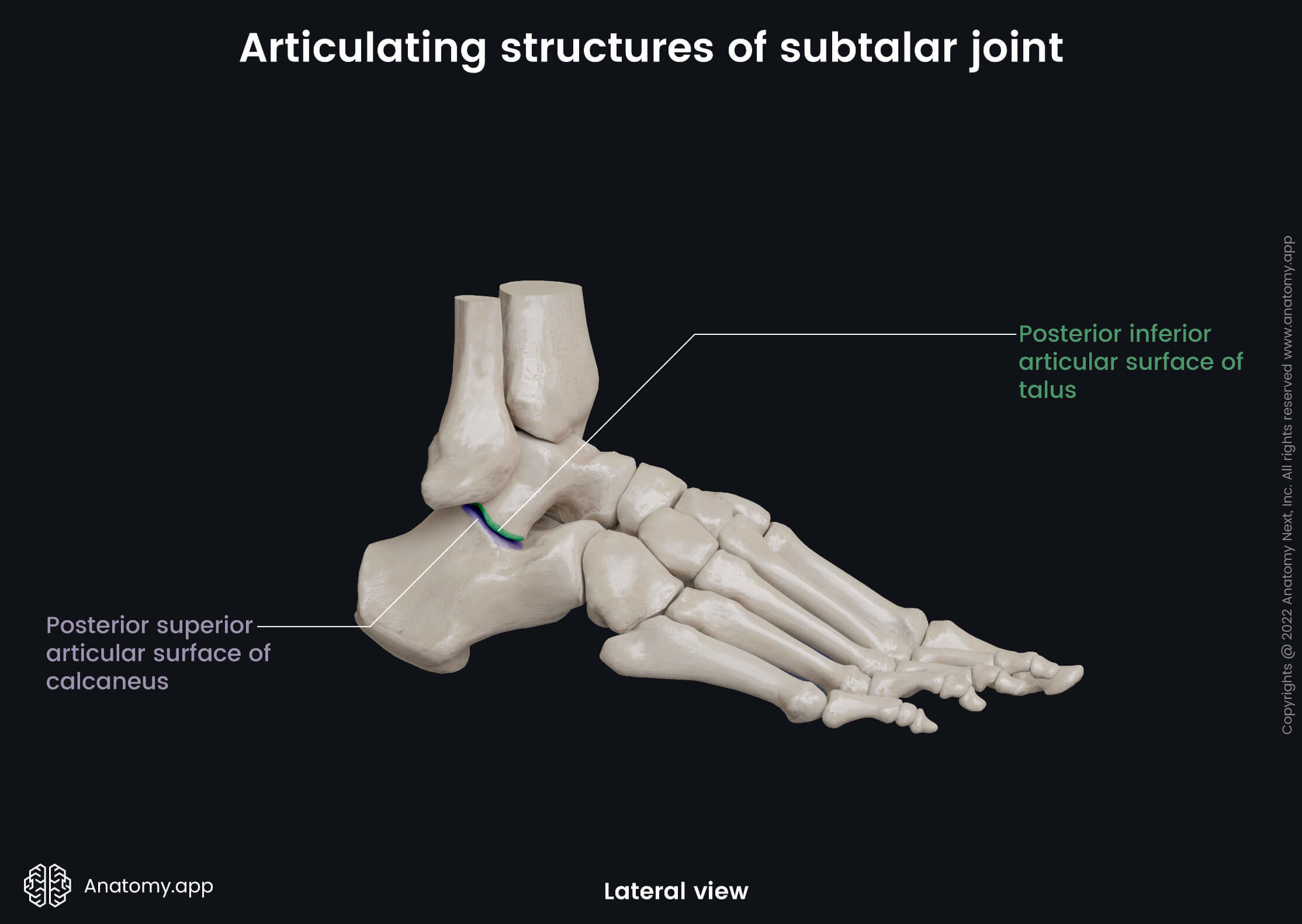
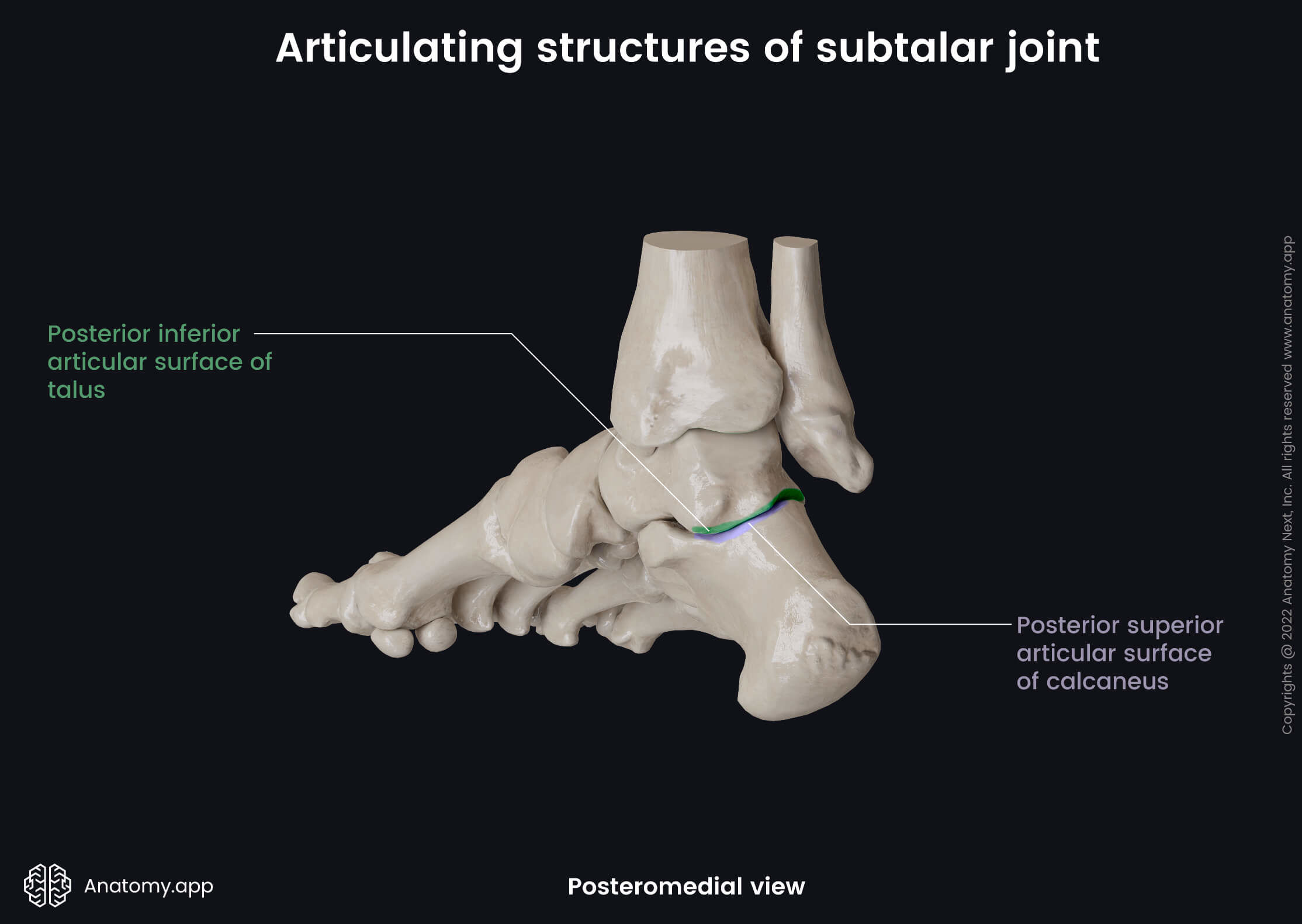
The articular surfaces of the subtalar joint include the following:
- Posterior inferior articular surface of the talus
- Posterior superior articular surface of the calcaneus
Joint capsule and ligaments
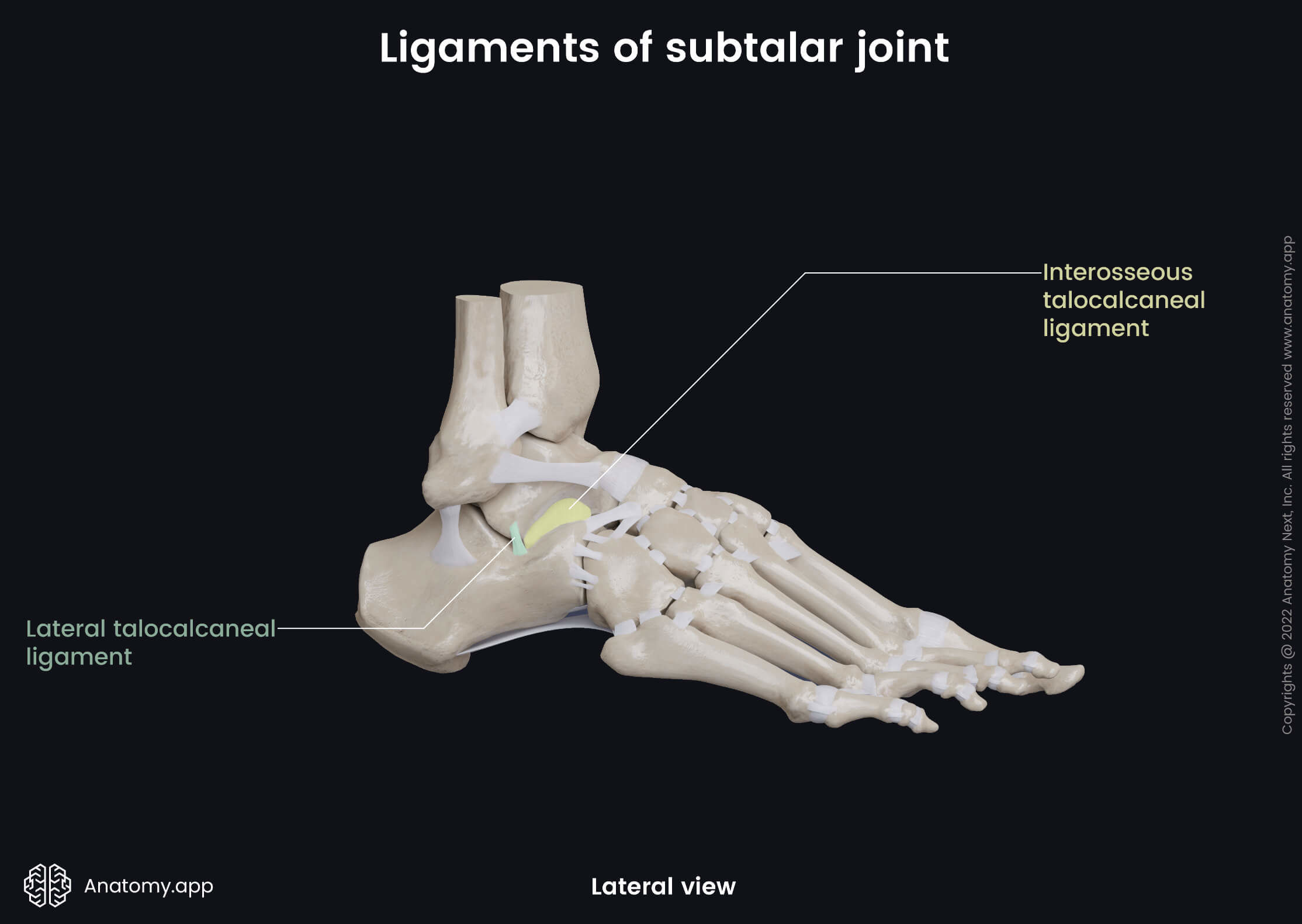
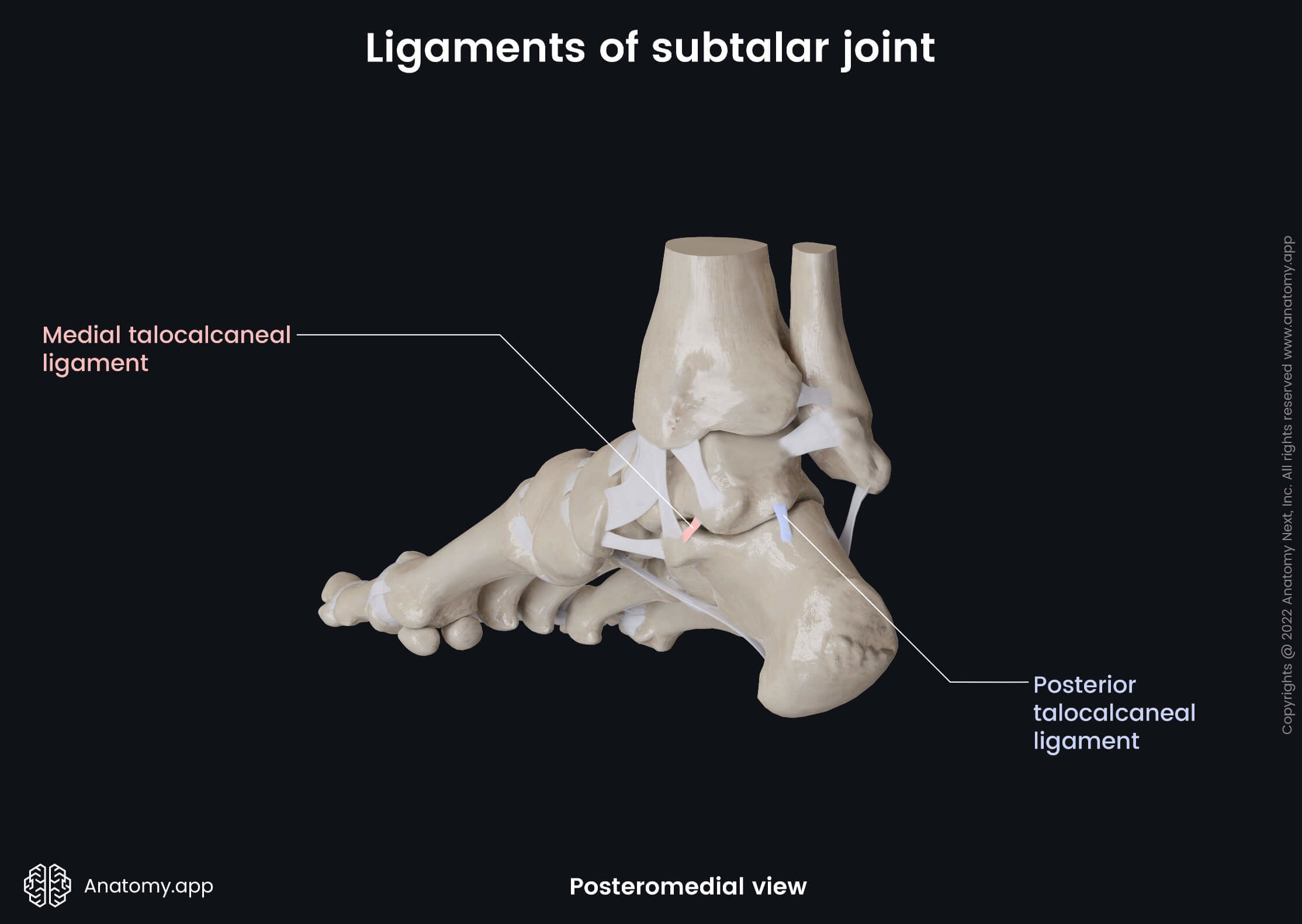
The bones that form the subtalar joint are enclosed by a fibrous joint capsule. The joint is stabilized by the following ligaments:
- Lateral talocalcaneal ligament - passes obliquely downward from the lateral talar process to the lateral calcaneal surface and calcaneofibular ligament;
- Medial talocalcaneal ligament - extends between the medial talar tubercle, the dorsal aspect of the sustentaculum tali and medial surface of the calcaneus;
- Posterior talocalcaneal ligament - stretches from the posterolateral tubercle of the talus to the superomedial area of the calcaneus;
- Interosseous talocalcaneal ligament - passes obliquely downward from the sulcus tali to the calcaneal sulcus.
Movements of subtalar joint
Movements of this joint happen simultaneously with the motions provided by the talocalcaneonavicular joint. This articulation permits the following movements:
- Supination and adduction of the foot
- Pronation and abduction of the foot