- Anatomical terminology
- Skeletal system
- Joints
- Muscles
- Heart
- Blood vessels
- Lymphatic system
- Nervous system
- Respiratory system
- Digestive system
- Urinary system
- Female reproductive system
- Male reproductive system
- Endocrine glands
- Eye
- Ear
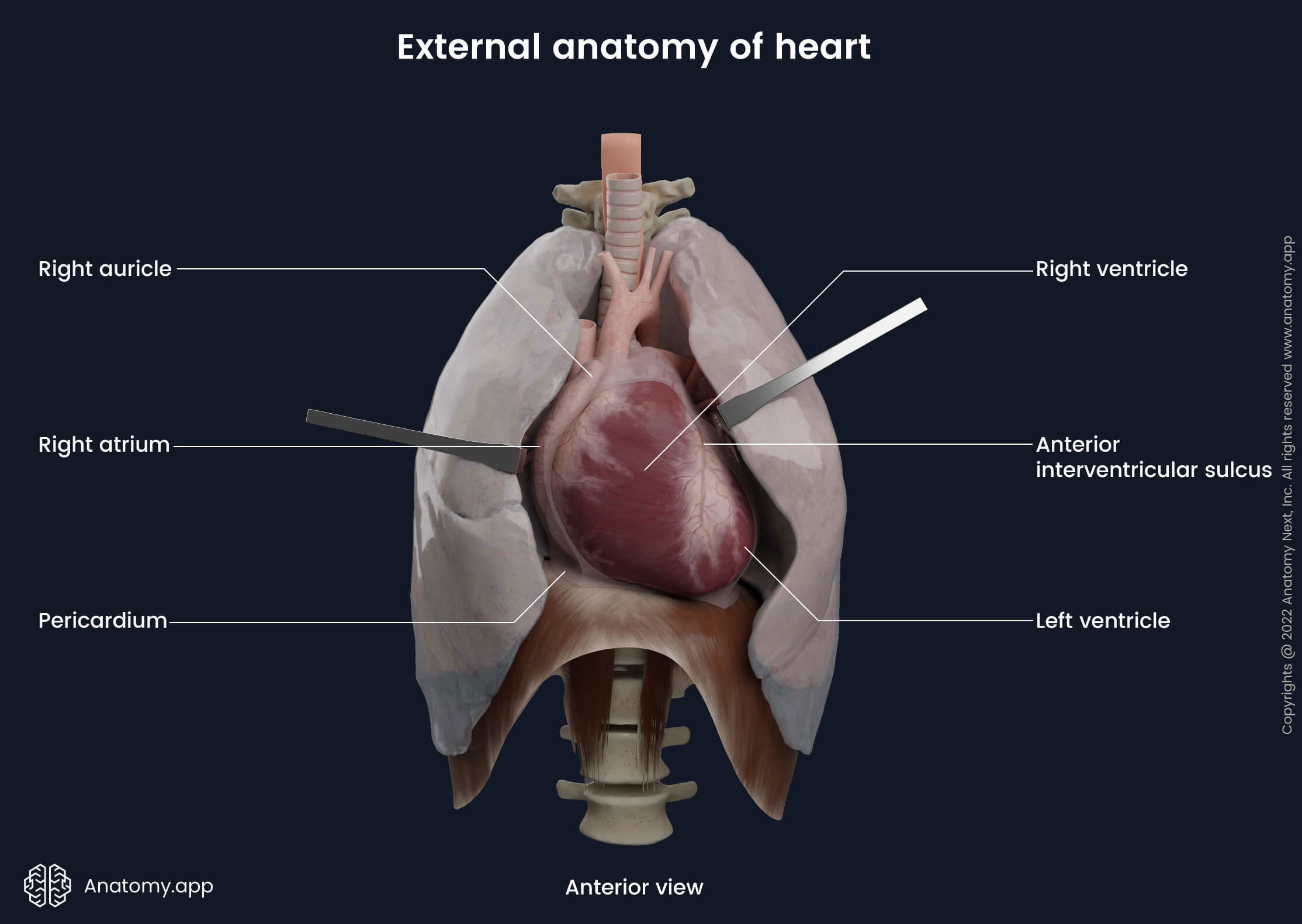
External heart anatomy
The heart has a cone or pyramid shape with its base projected upward, backward, and right. And the apex of the heart is directed forward, downward, and to the left. Approximately two-thirds of the heart is located on the left, while one-third on the right side from the midline of the body. The apex of the heart is typically located deep to the left 5th intercostal space and is formed by the left ventricle.
The longitudinal axis of the heart extends from the base to the apex. It goes from the top to the bottom, from right to left, and from back to the front. The heart is slightly rotated around the longitudinal axis to the left. The right side of the heart is located closer to the ventral surface of the body, while the left side - closer to the dorsal surface of the body.
The heart weighs around 200 to 400 grams (7 to 14 oz). Each day it beats about 100,000 times and pumps approximately 6 - 7,5 liters (1,6-2 gallons) of blood. The average heart rate is 60 to 90 heartbeats per minute. Externally, the heart has four borders and five surfaces formed by different internal parts. The external surfaces of the heart contain grooves - external sulci.
Heart borders
The heart has four borders separating the surfaces:
- Superior border - formed by the right and left atrium, auricles, and the great vessels;
- Right border - formed by the right atrium extending between the superior and inferior vena cava;
- Left border - created by the left ventricle and left auricle;
- Inferior border - formed by the left and right ventricles.
Position of heart
The superior border of the heart goes horizontally at the level of the 3rd rib cartilage, and the right one goes archwise down from the 3rd rib cartilage to the 5th rib cartilage. This border is located approximately 1,5 centimeters from the sternum on the right side. The inferior border goes obliquely down from the cartilage of the 5th rib on the right side to the apex of the heart on the left side.
The apex of the heart is projected in the fifth intercostal space. It is approximately 1,5 centimeters medially from the left midclavicular line. The left border goes obliquely upwards from the apex to the site where the cartilage of the third rib joins the bony part of the rib.
Heart surfaces
The heart has five surfaces:
- Diaphragmatic surface (inferiorly)
- Sternocostal surface (anteriorly)
- Right and left pulmonary surfaces (medially and laterally)
- Base of heart (posteriorly)
Diaphragmatic surface
The heart in its anatomical position rests on the diaphragmatic surface, which is a flat surface. It is faced inferiorly and lies on the respiratory diaphragm. This surface is formed by the right ventricle and a small part of the left ventricle separated by the posterior interventricular groove.
Sternocostal surface
The sternocostal (anterior) surface of the heart is convex. It is directed anteriorly and slightly to the left against the sternum and inner surfaces of the ribs. It consists mainly of the right ventricle, a part of the right atrium, and part of the left ventricle. On the sternocostal surface above the coronary sulcus are located two auricles - right and left. The right one is larger than the left one.
Pulmonary surfaces
The right and left pulmonary surfaces are broad, rounded, convex, and face the lungs. The left pulmonary surface is formed by the left ventricle and a part of the left atrium. And the right pulmonary surface consists of the right atrium.
Base of heart
The base of the heart is quadrilateral and oriented posteriorly. It consists of the left atrium, a small portion of the right atrium, and the proximal parts of the great veins (superior vena cava and inferior vena cava, as well as the pulmonary veins). The base of the heart is fixed posteriorly to the pericardial wall, opposite the bodies of the 4th/5th to 8th thoracic vertebrae (5th/6th to 9th when standing).
External sulci of heart
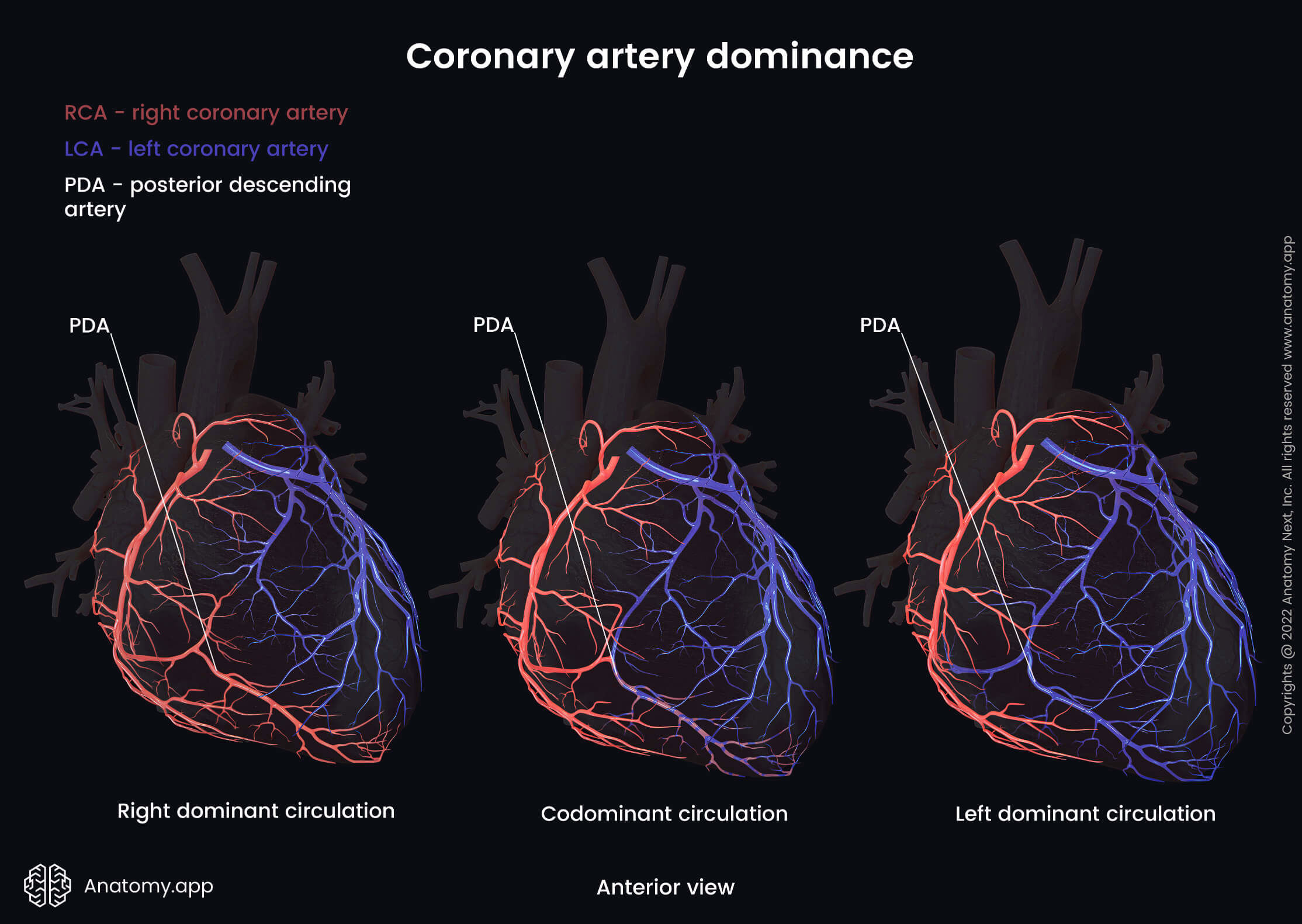
The internal partitions forming four chambers produce grooves on the external surface called external sulci, including the coronary sulcus and the anterior and posterior interventricular sulci. All of these external sulci are continuous with each other inferiorly, just to the right of the apex. The area on the lower backside of the heart where the coronary sulcus meets the posterior interventricular sulcus is called the cardiac crux or crux of the heart.
Coronary sulcus
The coronary sulcus is also known as the atrioventricular groove. It is a groove that circles the heart, marking the separation between the atria and the ventricles. It contains the right coronary artery, the small cardiac vein, the coronary sinus, and the circumflex branch of the left coronary artery.
Anterior and posterior interventricular sulci
The anterior and posterior interventricular sulci go in a vertical direction and mark the separation of both ventricles. The anterior interventricular sulcus is situated on the anterior surface (sternocostal surface). It contains the anterior interventricular artery and the great cardiac vein. The posterior interventricular sulcus is located on the diaphragmatic surface. It contains the posterior interventricular artery and the middle cardiac vein.
Anatomical relations of heart
Anteriorly to the heart is located the sternum and cartilages of the ribs, and the left lung. Posteriorly organs of the posterior mediastinum are situated, including the esophagus, descending thoracic aorta, azygos vein, hemiazygos vein and thoracic duct, as well as other blood vessels, nerves, and lymph nodes.
Below the heart - inferiorly - lies the central tendon of the respiratory diaphragm. Above or superiorly the large blood vessels of the heart and the bifurcation of the pulmonary trunk are located. The lateral sides of the heart are related to the mediastinal part of the parietal pleura and lungs.
Layers of heart wall
The wall of the heart is formed by three layers:
- Endocardium - the inner layer;
- Myocardium - the middle layer;
- Epicardium - the outer layer.
The heart with all three layers is covered by the pericardium.
Endocardium
The endocardium is the inner layer of the heart wall, and it lines all the internal surfaces of the heart cavities. It covers the pectinate and papillary muscles, trabeculae carneae, valves, and chordae tendineae.
The structure of the endocardium is similar throughout the heart. However, in the left atrium and ventricle, the endocardium is thicker than in the right atrium and ventricle. And the atrial endocardium is more prominent than the ventricular endocardium. Part of the endocardium covering the chordae tendinae is very thin.
All of the heart valves are derived from the endocardium, including:
- Valve of the inferior vena cava and valve of the coronary sinus in the right atrium;
- Right atrioventricular (three cusps) and pulmonary valves in the right ventricle;
- Left atrioventricular (two cusps) and aortic valves in the left ventricle.
At the openings of the arteries and veins, the endocardium passes into the walls of blood vessels.
Myocardium
The myocardium is a thick muscular layer between the endocardium and epicardium. It is formed by cardiac muscle fibers, also known as cardiomyocytes. The primary function of the myocardium is to contract and pump blood out of the heart to provide organs with blood. Cardiac muscles are characterized by rapid contractions that can not be controlled. The myocardial layer is different in the atria and ventricles.
In the atria, the myocardium has two layers: an outer circular layer that is common in both atria, and an inner longitudinal layer that is separate in each atrium. The longitudinal layer is formed by pectinate muscles.
In the ventricles, the myocardium is formed by three layers: outer and inner layers that are longitudinal and are common in both ventricles, and a middle layer that is circular and separate in each ventricle.
In the heart ventricles, the muscles of the inner layer form the papillary muscles and the trabeculae carneae. The outer longitudinal layer at the apex of the heart creates the vortex of the heart - the swirling arrangement of cardiac muscular fibers at the apex of the heart. At the mentioned site, the outer longitudinal layer transitions into the inner longitudinal layer. It happens vice versa as the inner longitudinal layer transitions into the outer longitudinal layer.
Epicardium
The epicardium is also called the visceral or inner layer of the serous pericardium. It is the outer serous layer of the heart. It is a thin, transparent layer composed of loose connective tissue, elastic fibers, and adipose tissue.
The epicardium covers not only the heart but also the beginnings of the great vessels - the ascending aorta, pulmonary trunk, superior vena cava, inferior vena cava, as well as the pulmonary veins before they return to the heart.
From these blood vessels, the epicardium further continues as the parietal layer of the serous pericardium. The epicardium protects the inner heart layers. It participates in the production of the pericardial fluid.
Pericardium
The heart is surrounded by a closed fibrous sac called the pericardium. The pericardium separates the heart from the surrounding organs. The pericardium contains two layers - an outer or fibrous and an inner or serous layer.
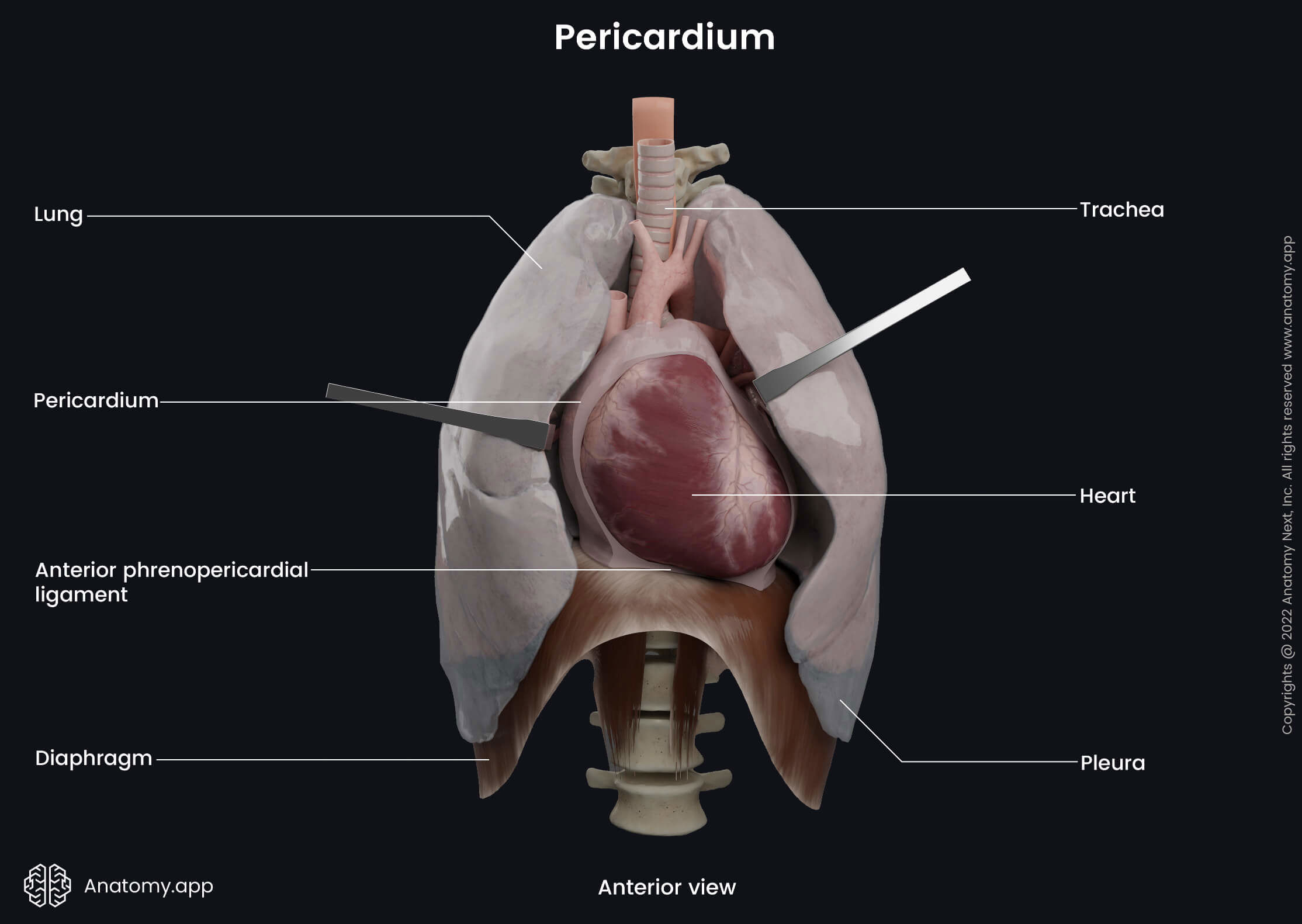
Fibrous pericardium
The outer or the fibrous layer of the pericardium forms a closed sac around the heart and separates the heart from the surrounding organs. It fuses with the respiratory diaphragm and pleura. The anterior surface of the pericardium connects with the inner surface of the sternum and ribs. The inferior surface of the pericardium fuses with the central tendon of the diaphragm and the anterior part of the muscular part of the diaphragm. The lateral surfaces fuse with the mediastinal pleura.
Serous pericardium
The serous layer of the pericardium has two lamina - the visceral and parietal lamina. The visceral lamina of the serous pericardium is also known as the epicardium. It covers the outer surface of the myocardium, and at the great blood vessels of the heart, it continues as the parietal lamina. The parietal lamina covers the heart and fuses together with the fibrous pericardium.
Pericardial cavity
The space between the parietal and visceral laminae of the serous pericardium is called the pericardial cavity. It contains a small amount of serous fluid (pericardial fluid) that decreases surface tension. This fluid also lubricates surfaces, allowing free movement of the heart during contractions. The pericardial cavity contains few recesses and two sinuses - transverse pericardial sinus and oblique pericardial sinus.
Neurovascular supply of heart
Arterial blood supply of heart
The arterial blood supply to the heart and heart muscles is provided by the left and right coronary arteries and their branches.
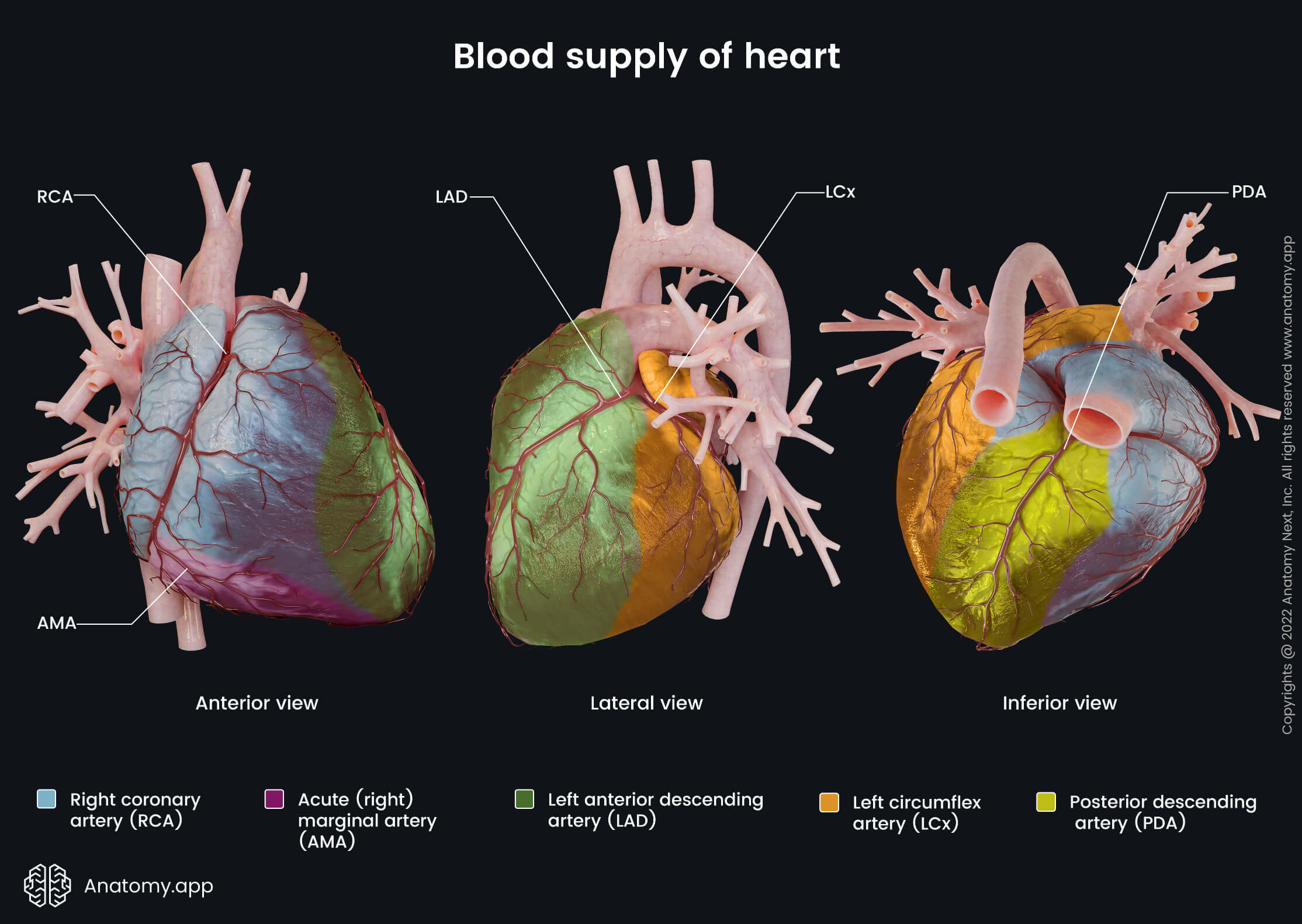
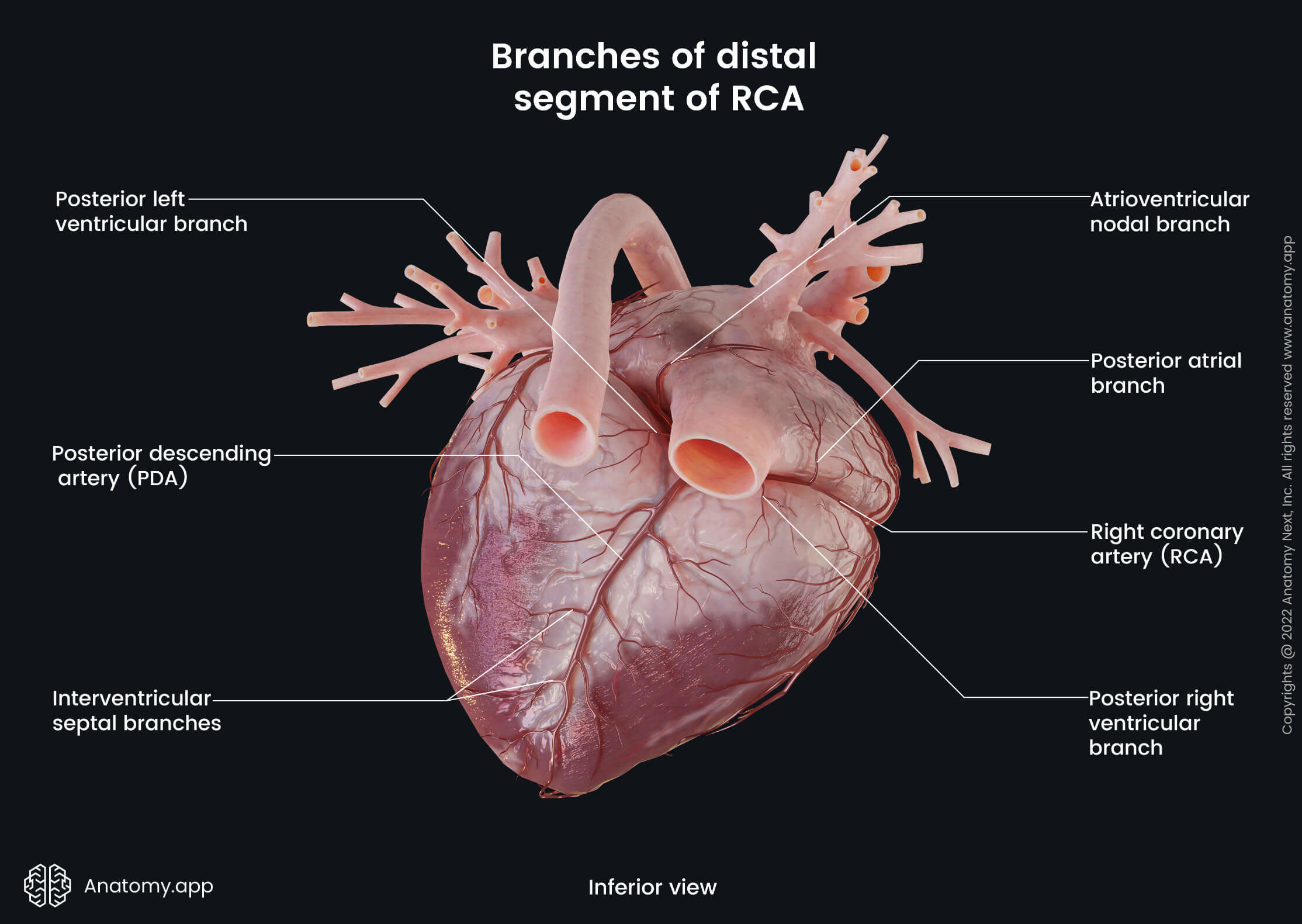
Right coronary artery
The right coronary artery has the following branches supplying the heart:
- Sinoatrial nodal branch
- Right marginal branch
- Atrioventricular nodal branch
- Posterior interventricular branch
All mentioned branches except the right marginal artery supply the right atrium and the right ventricle. The right marginal artery supplies the right ventricle and the apex of the heart. The posterior interventricular artery also supplies the interventricular septum.
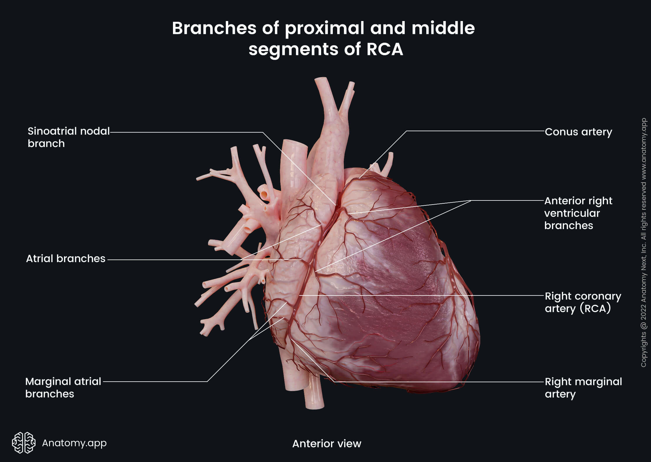
To sum up, the right coronary artery with its branches supply the right atrium and interatrial septum, sinoatrial and atrioventricular nodes, all posterior and partially anterior wall of the right ventricle, posterior third of the interventricular septum, all papillary muscles in the right ventricle, posterior papillary muscle in the left ventricle, a small part of the posterior wall of the left ventricle.
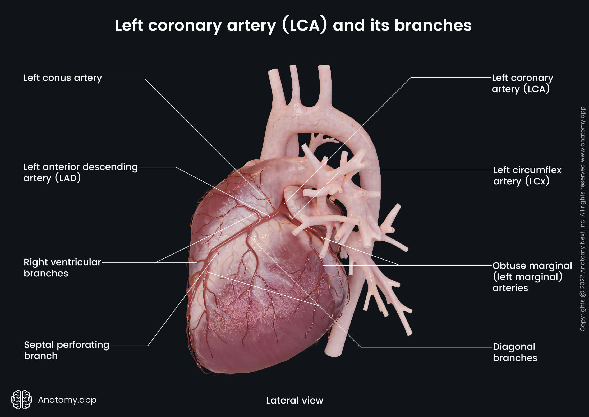
Left coronary artery
The left coronary artery has the following branches supplying the heart:
To sum up, the left coronary artery perfuses the left atrium and the left ventricle (except a small portion of the posterior wall), part of the anterior wall of the right ventricle, two anterior thirds of the interventricular septum, atrioventricular bundle, and anterior papillary muscles in the left ventricle.
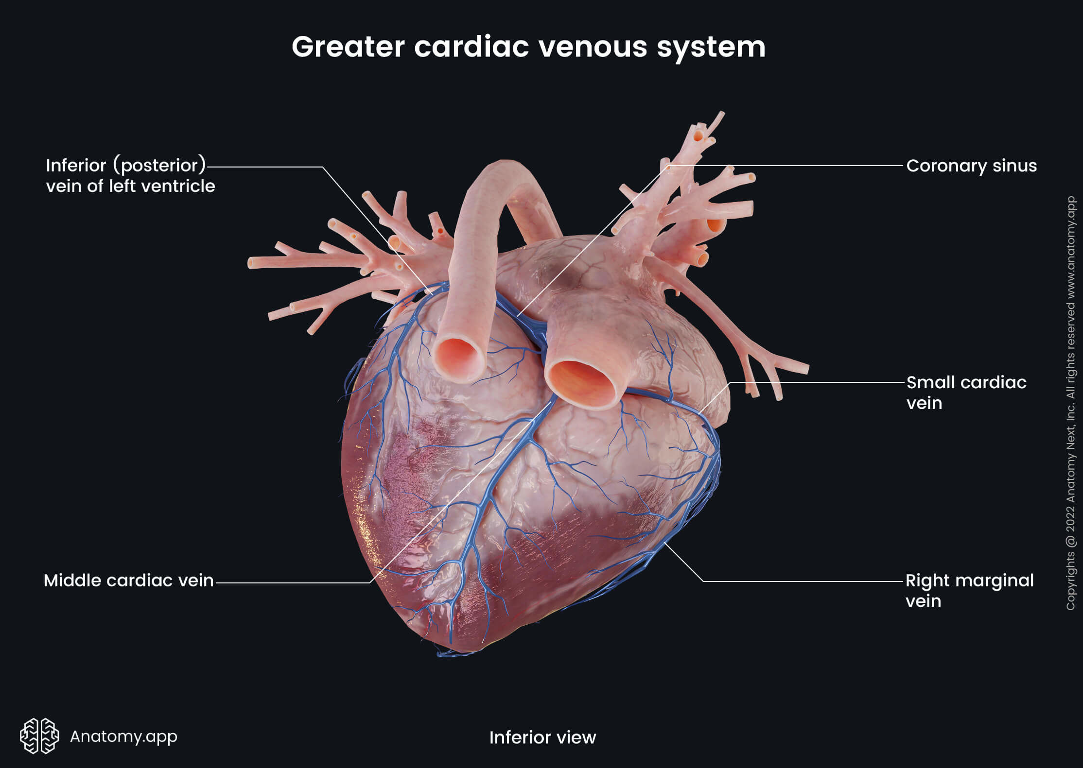
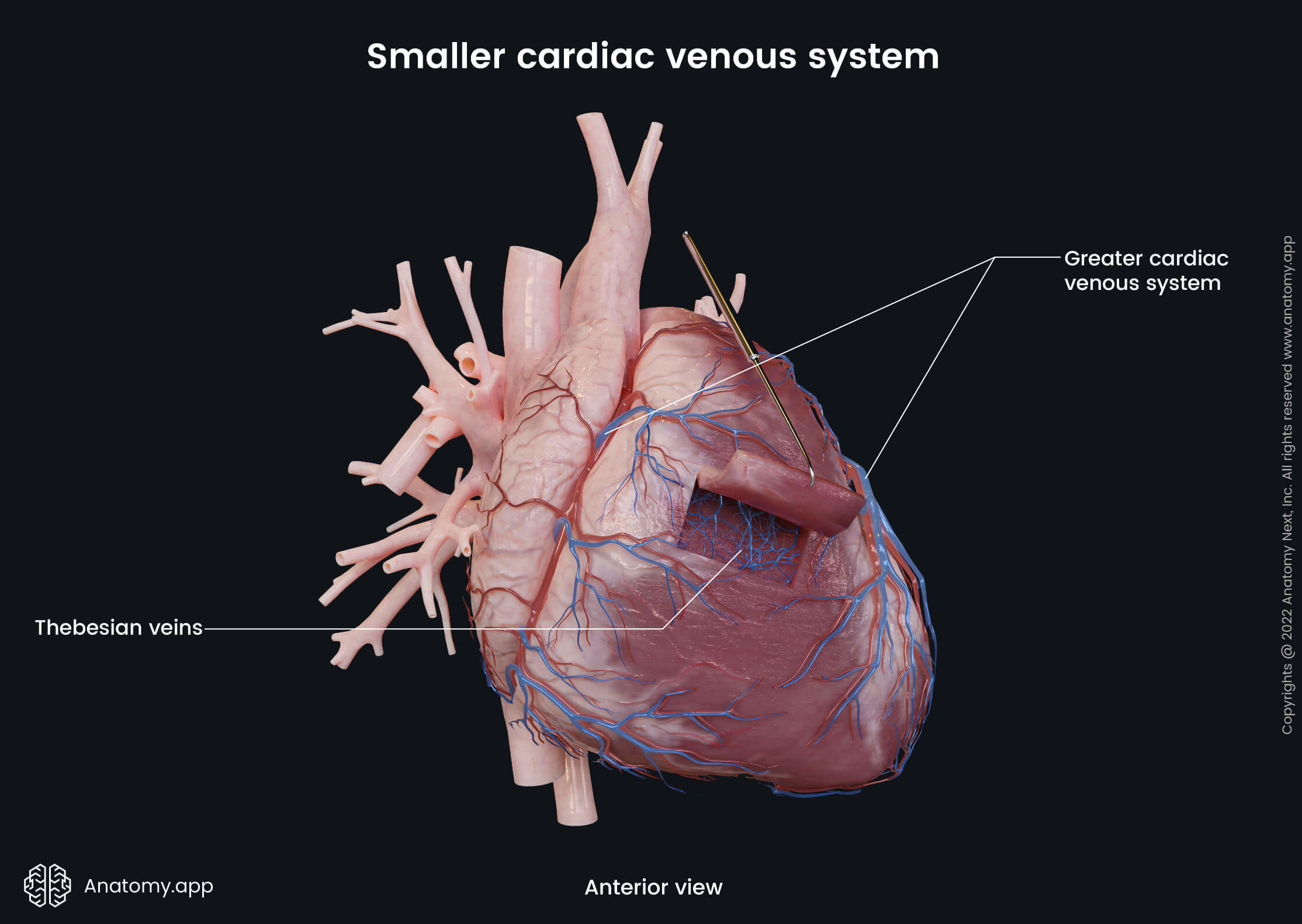
Venous drainage of heart
The coronary sinus provides venous drainage of the heart with its tributaries. The coronary sinus drains into the right atrium. The main tributary of the coronary sinus is the great cardiac vein arising at the apex of the heart. Besides the great cardiac vein, the middle and small cardiac veins also drain into the coronary sinus. The left marginal vein and left posterior ventricular veins also provide the drainage of the heart.


Lymphatic drainage of heart
Lymph is drained from the heart tissue via the subendocardial, myocardial lymph nodes and then via subepicardial plexus forming the right and left cardiac collecting trunks. The left trunk drains into the tracheobronchial lymph nodes, and the right trunk - into the brachiocephalic lymph nodes.
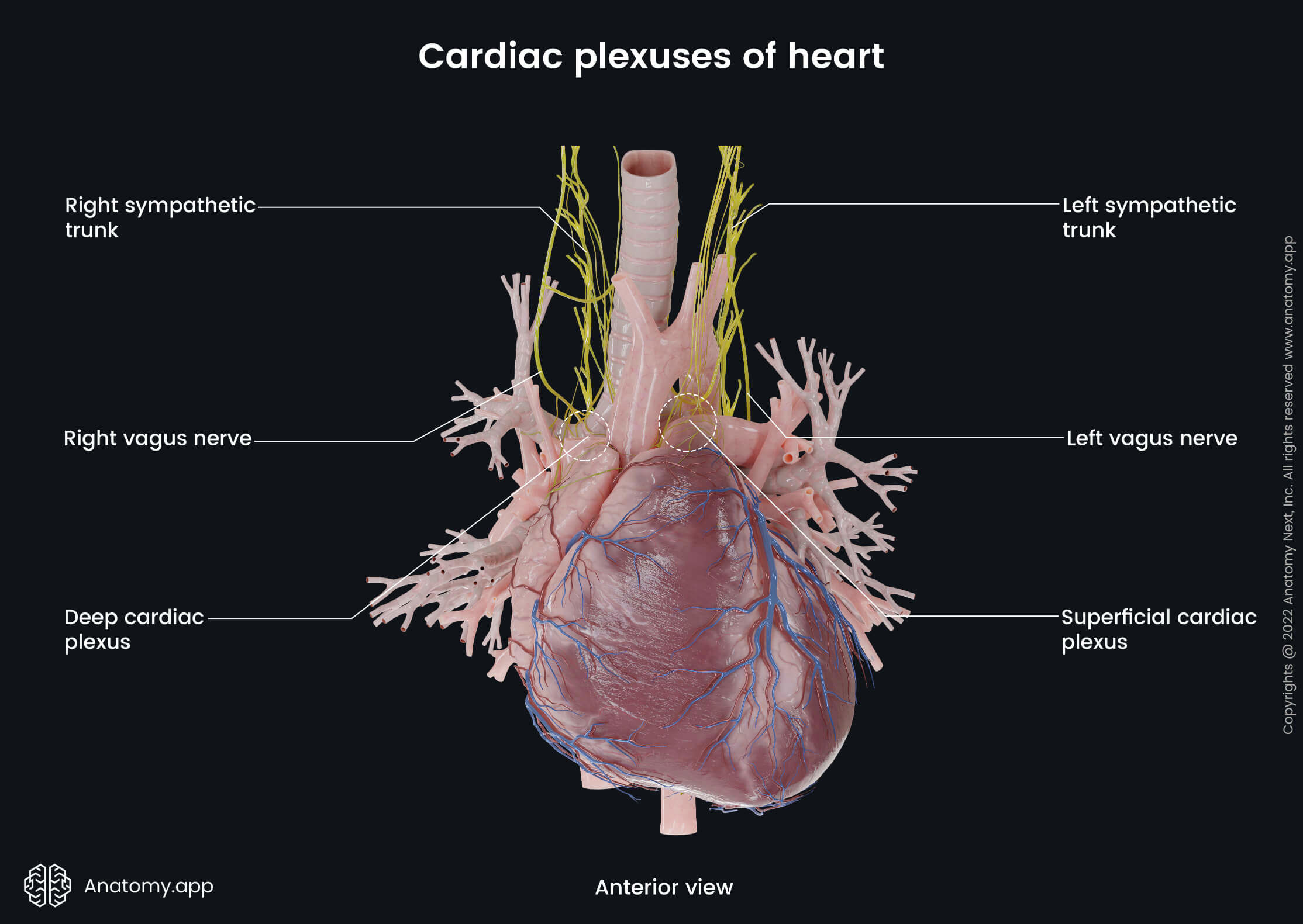
Innervation of heart
The heart is innervated by sympathetic and parasympathetic fibers from the autonomic nervous system, which form the cardiac plexus (contains fibers from both mentioned parts of the autonomic nervous system). Parasympathetic efferent and afferent fibers to this plexus are provided by the vagus nerve (CN X). Impulses of the parasympathetic fibers reduce heart rate and force of myocardial contraction, and also provide vasoconstriction of the coronary arteries. The vagus nerve gives off several cardiac branches such as thoracic cardiac, inferior and superior cervical cardiac branches.
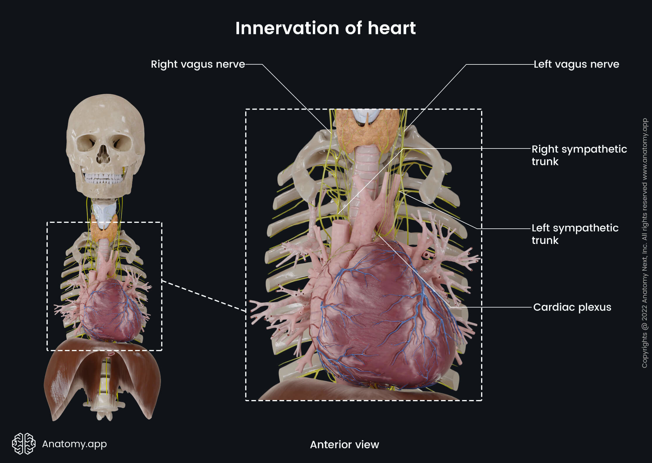
The sympathetic efferent innervation is provided by the sympathetic trunk via cardiac nerves of the lower cervical and upper thoracic ganglia. These nerves, in contrast to the parasympathetic nerves, increase heart rate and the force of contraction of the myocardium. The sympathetic afferent innervation is also provided by the lower cervical and upper thoracic ganglia. Sympathetic afferent fibers provide the pain sensation of the heart.
Anatomy.app
Contact information
- For questions regarding business inquiries. Please contact:
- info@anatomy.app