- Anatomical terminology
- Skeletal system
- Joints
- Muscles
- Heart
- Blood vessels
- Lymphatic system
- Nervous system
- Respiratory system
- Digestive system
- Urinary system
- Female reproductive system
- Male reproductive system
- Endocrine glands
- Eye
- Ear
Internal heart anatomy
Internally, the heart is divided into four chambers: right and left atria (singular: atrium) and right and left ventricles. The chambers are separated by septa containing subdivisions. The interatrial subdivision is the upper part, located between atria. The interventricular subdivision is the lower portion located between the ventricles. But the atrioventricular part is located between the atria and ventricles.
The heart consists of two pumps (right and left), each formed by an atrium and a ventricle separated by a valve. The right pump (consisting of right atrium and right ventricle) receives deoxygenated blood and pumps it into the lungs. And the left pump (left atrium and left ventricle) receives oxygenated blood from the lungs and sends it to the body. The atria receive blood, thus, have relatively thin walls, while the ventricles have reasonably thick walls, as they pump blood out of the heart.
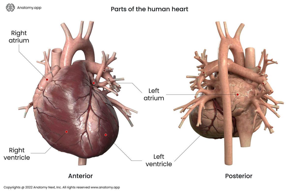
Right atrium
The right atrium of the heart is situated in the superior right corner of the heart above the right ventricle. The systemic circulation ends in the right atrium. The deoxygenated blood enters it through the inferior and superior vena cava and the coronary sinus.
Walls of right atrium
The right atrium has a cuboid shape. Therefore it has six walls:
- Superior
- Inferior
- Anterior
- Posterior
- Lateral
- Medial
Between the medial and posterior walls below the depression known as fossa ovalis is located a small opening called the opening of the coronary sinus. Through this opening, the coronary sinus opens to the right atrium. The coronary sinus collects venous blood from the heart muscle. It is protected by a semicircular fold of the lining membrane, called the valve of the coronary sinus (also known as the Thebesian valve).
The superior vena cava opens to the superior wall of the right atrium with an aperture called opening of the superior vena cava. On the inferior wall of the right atrium the right atrioventricular orifice is found. It is an oval opening between the right atrium and the right ventricle.
On the anterior wall is placed the right auricle. The primary function of the right auricle is to increase the volume of the right atrium. The muscular layer of the heart creates parallel ridges in the anterior wall and the auricle. They are called the pectineal muscles. On the posterior wall the opening of the inferior vena cava is located where inferior vena cava opens to the right atrium.
Along the inferior margin of the opening of the inferior vena cava extends a semilunar valve called the valve of the inferior vena cava. The valve functions in the fetus until birth. It directs blood from the right atrium through the foramen ovale to the left atrium. The enlarged posterior part of the right atrium receiving blood directly from both inferior and superior venae cavae is called the sinus of the venae cavae.
On the lateral wall of the right atrium the pectineal muscles are located. The medial wall is formed by the interatrial septum. In the interatrial septum lies an oval fibrous depression called the fossa ovalis covering the foramen ovale during fetal development. Around the fossa ovalis is an oval margin named the annulus ovalis or limbus of the fossa ovalis. Small openings for the smallest cardiac veins are also located on the medial wall of the right atrium.
Right ventricle
The right ventricle is one of the heart chambers located in the inferior right portion of the heart under the right atrium and opposite to the left ventricle. The right ventricle contains deoxygenated blood. The primary function of the right ventricle is to pump blood up through the pulmonary valve and trunk into the lungs, thus providing pulmonary circulation.
The right ventricle has a pyramidal shape with the base directed upwards and the apex - downwards. The medial wall of the right ventricle is formed by the interventricular septum. The inferior wall lies close to the central tendon of the diaphragm. The anterior wall of the right ventricle is directed towards the inner surfaces of the sternum and ribs.
The base of the right ventricle contains the right atrioventricular orifice and the opening of the pulmonary trunk. The part of the right ventricle that leads to the opening of the pulmonary trunk is called the infundibulum (or conus arteriosus). It is a conical extension formed from the upper and left angles of the right ventricle. The inner surface of the right ventricle is smooth only in the infundibulum. Elsewhere the muscular layer of the heart creates papillary muscles and irregular muscular columns called trabeculae carneae.
Right atrioventricular orifice and tricuspid valve
Venous blood from the right atrium enters the right ventricle through the right atrioventricular orifice. Around the right atrioventricular orifice is the tricuspid valve. The tricuspid valve has three leaflets - the anterior, posterior, and septal leaflets. Around the orifice is a fibrous ring called the fibrous annulus.
The right atrioventricular valve opens during atrial systole. It allows deoxygenated blood from the right atrium to flow into the right ventricle. The valve closes during the ventricular systole. The primary function of the tricuspid valve is to prevent the backflow of the blood from the ventricles into the atria.
There are three papillary muscles in the right ventricle. Leaflets are connected with chordae tendineae. The chordae tendineae arise from one leaflet and insert into two adjacent papillary muscles. The chordae tendineae prevent the valve leaflets from prolapsing into the right atrium. The papillary muscles fix the tricuspid valve, but trabeculae carneae prevents blood swirling.
Opening of pulmonary trunk and valve
The exit opening from the right ventricle is the opening of the pulmonary trunk. Deoxygenated blood enters the pulmonary trunk through this opening. The trunk divides into two pulmonary arteries - right and left pulmonary arteries. The opening is located behind the left sternocostal joint of the third rib.
Around the opening of the pulmonary trunk is the pulmonary valve, which is a semilunar valve. It has three semilunar cusps: anterior left and right. It is attached to a fibrous annulus located around the opening.
During ventricular systole, the pressure in the ventricle increases as the walls shrink. Valves are pressed against the inner surface of the pulmonary trunk, and the blood flows through it. As the pressure in the right ventricle decreases, the valve closes.
Left atrium
The left atrium is located in the superior left corner, above the left ventricle, and opposite to the right atrium. The pulmonary circulation ends in the left atrium, as oxygenated blood from the lungs enter it through the pulmonary veins.
The left atrium has a cuboid shape. It is thicker but smaller in volume than the right atrium. The medial wall of the left atrium is formed by the interatrial septum, which separates the left and right atria.
On the anterior wall of the left atrium lies the left auricle, a flap of the heart wall. The left auricle has an irregular shape with many tiny ridges created by the pectinate muscles. The primary function of the left auricle is to increase the volume of the left atrium. Elsewhere, the inner surface of the left atrium is smooth.
Openings of left atrium
There are five openings found in the walls of the left atrium. These include four openings for the pulmonary veins and one opening called left atrioventricular orifice. The four openings for the pulmonary veins are located on the superior and posterior walls of the left atrium. The pulmonary veins carry oxygenated blood from the lungs to the left atrium. The left atrioventricular orifice is found on the inferior wall of the left atrium. It carries oxygenated blood to the left ventricle.
Left ventricle
The left ventricle is one of the heart chambers located in the lower-left portion of the heart below the left atrium, opposite to the right ventricle. The primary function of the left ventricle is to pump blood into the aorta, providing systemic circulation.
The left ventricle has a cone shape with a base directed upward, while the apex is directed inferiorly. There are two openings found on the base of the left ventricle - the left atrioventricular orifice and aortic orifice.
There are fibrous rings and valves around both openings. The anterosuperior portion of the left ventricle below the aortic orifice is called the aortic vestibule. The inner surface of the heart is smooth at the aortic vestibule. Elsewhere the inner surface is covered by papillary muscles and trabeculae carneae.
Left atrioventricular orifice and mitral valve
Around the left atrioventricular orifice is the left atrioventricular valve (also called the mitral valve, bicuspid valve). The mitral valve has two cusps - the anterior and posterior. Between both cusps are smaller ones called the commissural cusps. The opening of the mitral valve is surrounded by a fibrous ring called the mitral annulus.
The chordae tendineae are inelastic tendons attached to the cusps and the papillary muscles within the left ventricle. The chordae tendineae prevent the valve leaflets from prolapsing into the left atrium.
The left atrioventricular valve opens during atrial systole, allowing arterial blood from the left atrium to enter the left ventricle. The valve closes during the ventricular systole.
Aortic orifice and aortic valve
The aortic orifice is guarded by the semilunar aortic valve. It is made of three cusps - left, right and posterior. The aortic orifice is surrounded by a fibrous ring called the annulus fibrosus, which fixes the aortic valve. The aortic orifice is located in the midline behind the sternum at the 3rd intercostal level.
Anatomy.app
Contact information
- For questions regarding business inquiries. Please contact:
- info@anatomy.app