Anatomy.app Content News in January and February

Welcome back to our Content News blog series, and actually…Happy New Year! We know it’s been a while since our last blog post was launched, and you've probably been looking for our monthly Content News articles. We have some really good excuses why there weren't any updates from us. Well, as always, we are thinking of you and have been working hard to deliver fresh, relevant, and high-quality content to you.
In the first two months of the year, we’ve created plenty of new 3D models, videos, illustrations, and animations, making Anatomy.app even more engaging and insightful. While we’ve been busy behind the scenes, we’re finally ready to share our latest updates with you. But you've probably already noticed some pretty amazing changes and additions, haven’t you?
As the spring is coming closer, Anatomy.app is also starting to show some never-before-seen flower buds that will soon be blooming in different beautiful colors. Stay tuned for new updates, and believe us, they will blow you away!
1. 3D Anatomy: New and Revised 3D Models
We are still very much into details and clear understanding of the subject at the same time. And it is what you guys love the most about our 3D models. Keeping this in mind, at the end of 2024 and the beginning of 2025, we added some new models to two of our 3D Anatomy articles - the Urinary system article and the Skull topography article.
Now, the 3D Skull topography article features not just one but various cross-sections of the skull. And you can clearly see all the paranasal sinuses in these new models. Finding it difficult to visualize the ethmoidal air cells? Well, we’ve got you covered.
In our 3D Skull topography article, you can zoom in and out of each ethmoidal air cell - how cool is that? Whether you want to take a closer look at the anterior or posterior ethmoidal air cell, just go to our 3D Skull topography article. All the ethmoidal air cells are there, patiently waiting for you to dive deeper into this study field!
Besides these new additions, one more article received a makeover. We have recently freshened up our 3D Male reproductive system article. In this article, we revised and updated some of the old slides and 3D models, as well as added some new slides containing fresh, just out of the oven 3D models.
Did you know that testicles are covered by several tissue layers that are formed during the descent of the testicles? You didn’t? Then head just straight to the revised and updated 3D Male reproductive system article: https://anatomy.app/article/male-reproductive-system/internal-and-external-male-genital-organs
Here is also a link to the 3D Skull topography article: https://anatomy.app/article/skull-topography/paranasal-sinuses-part-1
And don’t forget to explore the new 3D models in the 3D Urinary system article: https://anatomy.app/article/urinary-system
2. Media Library: New Animated 3D Materials
We also continued to expand our Media Library and added several new animated 3D materials. For some of you, biomechanics was once a nightmare to understand, but not anymore. Watching our animated 3D materials is like watching your favorite movie - just don’t forget to grab some popcorn.
We did these fresh animations on various topics, but most of them focus on the movements performed at the joints of the lower limb. However, there is one particular animation we want to spotlight - it is the nutation and counternutation animation.
Nutation and counternutation are movements that occur in the sacroiliac joints (SIJs), and they are important not only in the context of posture and walking but also in childbirth. We are sure that those interested in obstetric anatomy will greatly appreciate this new addition, as it clearly shows how the sacrum and hip bones interact during these movements. You can also easily track the motion of the coccyx.
So, here is a full list of our freshly added animated 3D materials:
- Circumduction of the head and neck
- Horizontal flexion and extension (horizontal adduction and abduction) of the upper limb
- Combined flexion and adduction, and extension and adduction at the hip joint
- Nutation and counternutation
- Inversion and eversion of the foot
- Abduction and adduction of the foot
- Flexion and extension at the interphalangeal joints
- Flexion and extension at the metatarsophalangeal joints
- Abduction and adduction at the metatarsophalangeal joints
Don’t waste a minute! Explore the new animations now: https://anatomy.app/media?categoryType=regions&mediaType=animatedModel
3. Encyclopedia: a Ton of Fresh Illustrations
At the start of this year, our Media Library and Encyclopedia got a fresh infusion of stunning new illustrations! As mentioned earlier, we updated our 3D Urinary system and 3D Skull topography articles, and illustrations, of course, came along with these new additions. But that's not all!
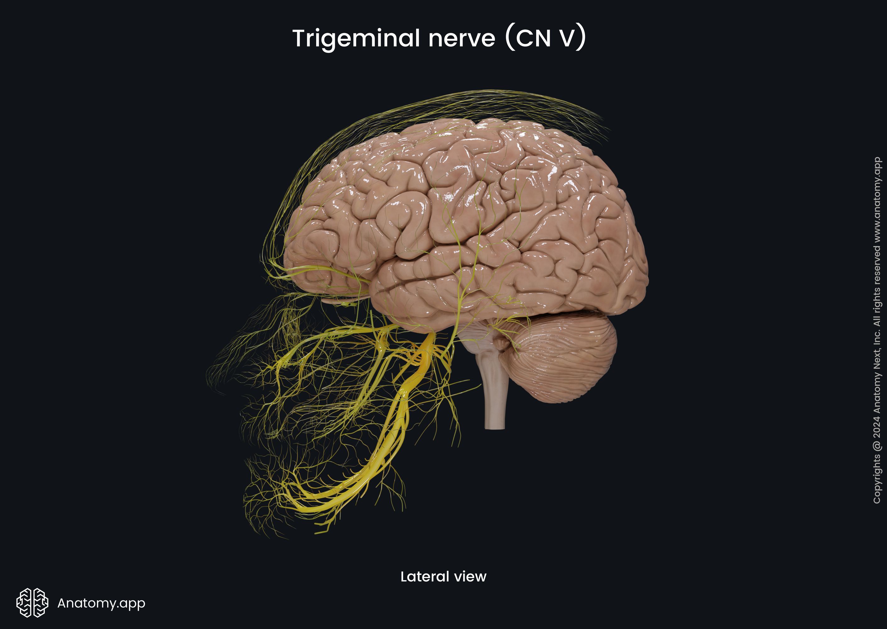
One of the most fascinating illustrations we created was on the fifth cranial nerve - the trigeminal nerve - and its branches. In these new visuals, you can undoubtedly see how important the trigeminal nerve is when it comes to the innervation of the structures found within the head. This nerve has larger and smaller branches everywhere.
From the intricate neural connections to the hidden chambers of the paranasal sinuses, this latest batch of illustrations brings anatomy to life with mind-blowing detail and clarity. Whether you're more fascinated by the digestive, urinary, or skeletal systems, we've got something for all of you.
Here’s a complete list of the articles that received new visuals:
Step into our illustration world here: https://anatomy.app/media?categoryType=regions&mediaType=image
4. Media Library: New Videos
Not only did our Media Library receive new illustrations and animations, but we also enriched it with a treasure trove of fascinating new videos. Our latest video collection covers such study fields as the skull, joints, vascular system, and obstetric anatomy. These additions not only explore the anatomy of different body structures but also dig deeper into the biomechanics and movements that define how we function.
And now, we would like to highlight two of our new videos. But first, you need an introduction. We recently added a stunning 3D model to our Media Library - a 3D female pelvis. To complement this, we made two comparison videos showcasing both pelvises we had. In the latest videos, you can clearly see the differences between them.
When watching the videos, keep in mind that just as people vary in size and shape, so do pelvises - there are variations. Overall, there are four main types of pelvises and the aim of our videos was not to represent pelvic types but simply to highlight the differences between the two pelvises we had.
Our 3D models are crafted from real scans of actual individuals. And we had two pelvises - one of which belonged to a female and the other to a male. The pelvises in our videos represent just two examples. It’s important to note that not all individuals will have these patterns exactly. Sometimes, only a few of these characteristics are present, or they may be only slightly pronounced.
Here’s a sneak peek at what’s waiting for you in our Media Library:
- Exploring the skull: sagittal section (with the brain included)
- Skull openings
- Biomechanics of the head and neck: circumduction
- Biomechanics of the wrist: circumduction
- Male vs. female pelvis
- Exploring differences: male and female pelvis comparison
- Biomechanics of the sacroiliac joints: nutation and counternutation
- Biomechanics of the hip joint: combined flexion and adduction and extension and adduction
- Biomechanics of the foot: inversion and eversion
- Overview of the blood vessels of the head and neck
- Blood vessels supplying the male urinary bladder and urethra
These fresh videos in easy-to-follow format are designed to provide a deeper and much clearer understanding of complex anatomical concepts. Whether you’re a medical student, a healthcare professional, or an anatomy enthusiast, we are sure these new additions will give you new insights and clarity on various aspects of human anatomy.
Watch our new videos now and take your anatomy expertise to the next level: https://anatomy.app/media?mediaType=video
Final Note
As we wrap up this year’s first edition of the Content News, we hope you’re just as excited and happy as we are about these latest updates. And we are even more excited to continue providing you with insightful and engaging content to make your educational journey more enjoyable, understandable and accessible. We hope you love anatomy just as much as we do!
So, keep your curiosity and passion for anatomy strong. Whether you’re exploring our new 3D models, diving into biomechanics with the help of our animated 3D materials, or watching our videos, there’s plenty of new content to discover. And trust us - this is just the beginning of what’s coming in 2025! Stay tuned, keep learning and see you in the next month’s update!
Until next time,
The Anatomy.app Team!🧠
Related blogs:
Anatomy.app
Contact information
- For questions regarding business inquiries. Please contact:
- info@anatomy.app