- Anatomical terminology
- Skeletal system
- Joints
- Muscles
- Heart
- Blood vessels
- Lymphatic system
- Nervous system
- Respiratory system
- Digestive system
- Urinary system
- Female reproductive system
- Male reproductive system
- Endocrine glands
- Eye
- Ear
Spleen
The spleen (Latin: splen s. lien) is a fist-sized and oval-shaped purplish secondary lymphoid organ and the largest organ of the lymphatic system. It has an essential role in immune surveillance and blood filtration. Overall, the spleen is the organ where various antigens and senescent and damaged red blood cells are filtered, but lymphocytes mature and proliferate.
Spleen anatomy
The spleen is a spongy, soft and very densely vascularized secondary lymphoid organ that is located posterolaterally in the left hypochondriac region of the abdomen. Because of its good vascularization, it appears reddish purple.
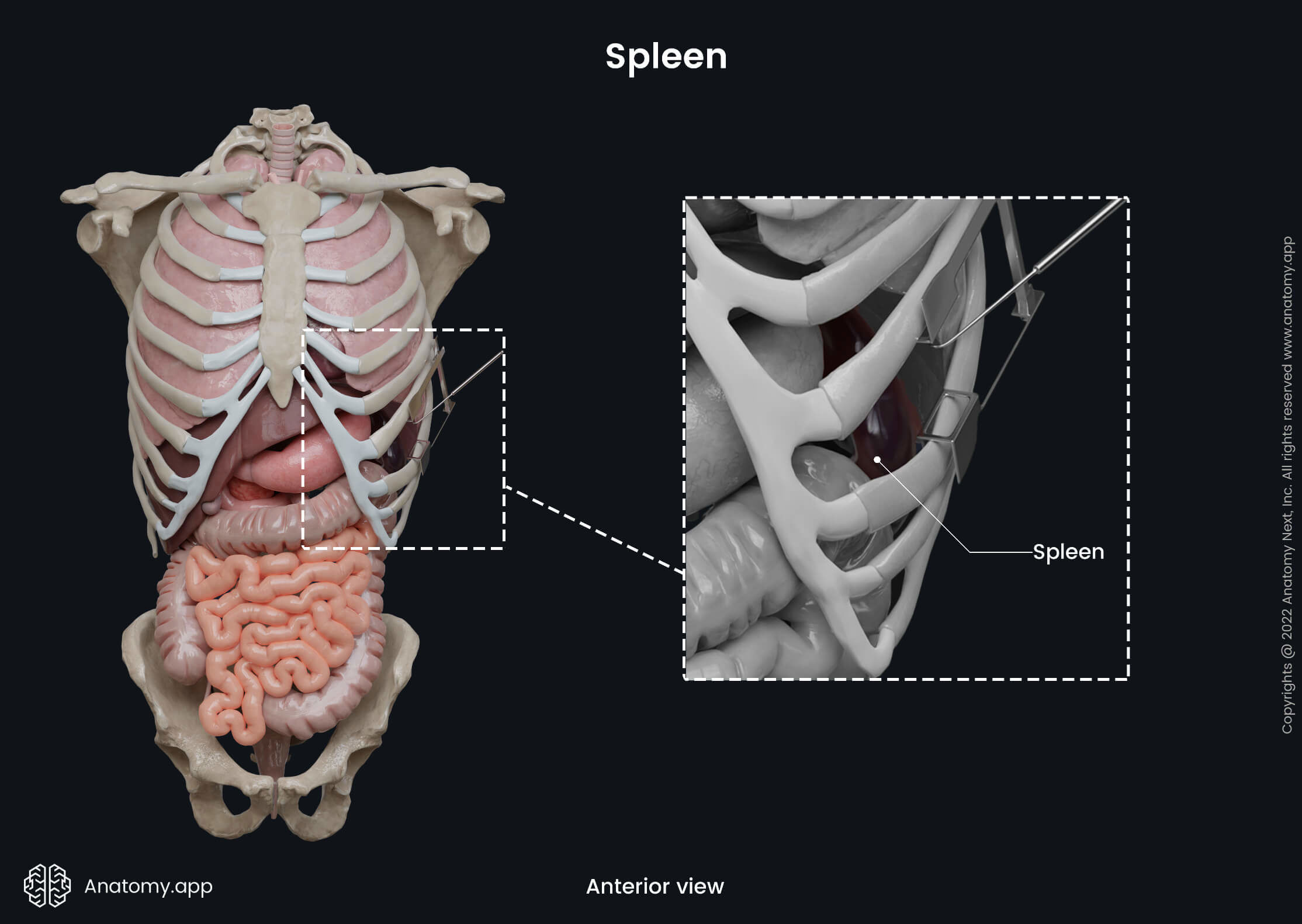
The spleen is found between the fundus of the stomach and the left hemidiaphragm at the level of ribs nine to eleven. It is classified as an intraperitoneal organ, meaning the peritoneum surrounds it from all sides except its hilum.
Note: The hilum of the spleen is a site where nerves, blood and lymphatic vessels leave and enter the spleen. It is found at the central aspect of the visceral surface of the spleen.
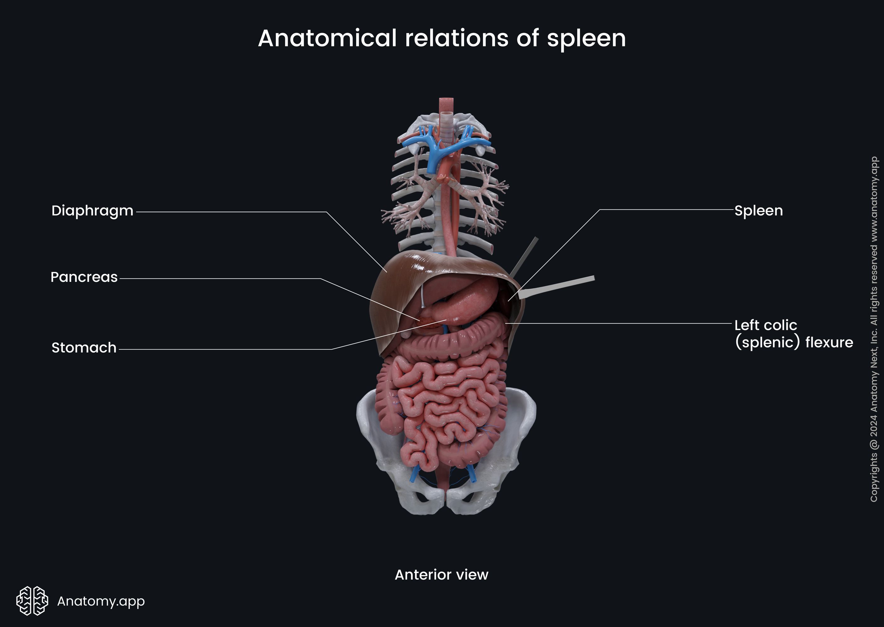
On the medial side of the spleen lies the left kidney, as well as adjacent to its medial aspect is the tail of the pancreas. In contrast, lateral to the spleen is the rib cage and muscles of the anterolateral abdominal wall. Superior to the spleen is the diaphragm, while inferior is the left colic flexure. Hence, the left colic flexure is also called the splenic flexure.
The spleen varies in shape and size depending on the gender, weight and height of a person. However, usually, it is somewhat oval-shaped and fist-sized. On average, it is 3.94 - 5.51 inches (10 - 14 centimeters) long, while its width varies from 2.76 to 3.54 inches (7 - 9 centimeters). The thickness of the spleen is typically 1.18 - 1.97 inches (3 - 5 centimeters). Usually, the spleen weighs around 4.9 - 7 ounces (140 - 200 grams).
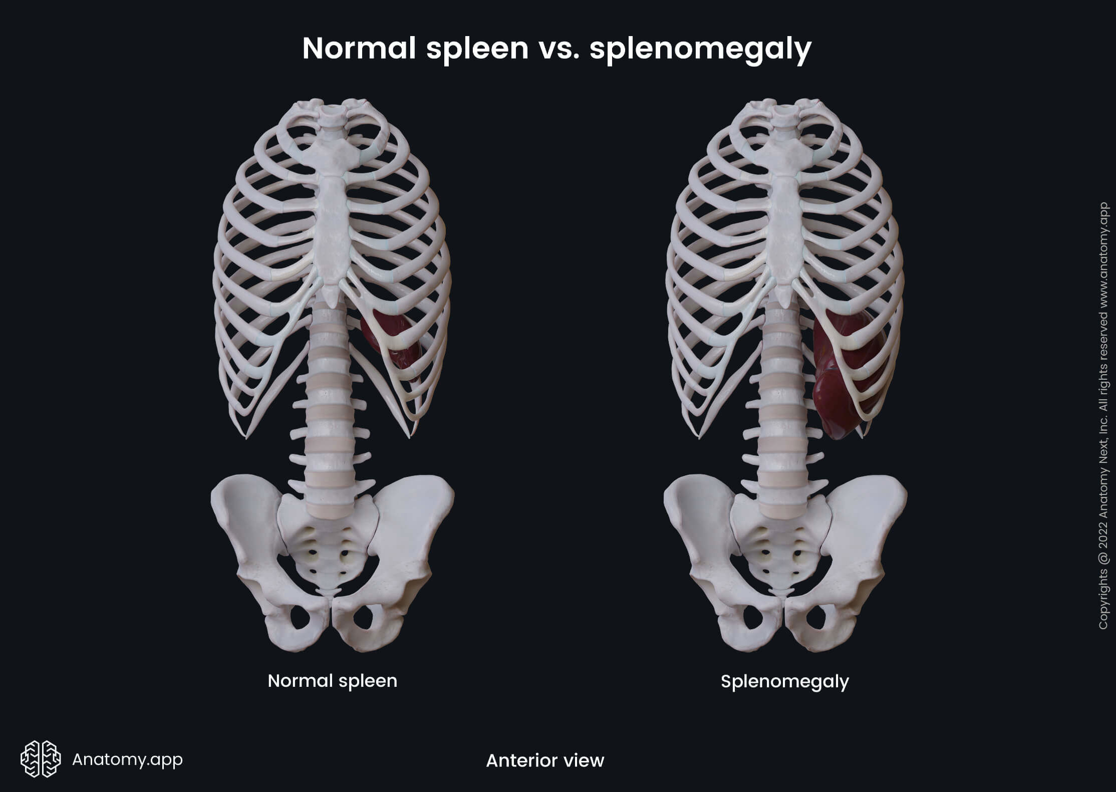
Typically, the spleen is not palpable in a healthy individual during physical examination, as it does not extend beyond the left costal arch. Doctors can seldom palpate it at the left costal arch through the anterolateral abdominal wall. An enlarged spleen is called splenomegaly, and in the case of this condition, it expands below the left costal arch, where it is easily palpable.
Overall, the spleen has two surfaces, three borders, and two extremities described below. Also, the spleen is surrounded by a connective tissue capsule and is connected to the adjacent internal organs and structures by several ligaments.
Spleen surfaces
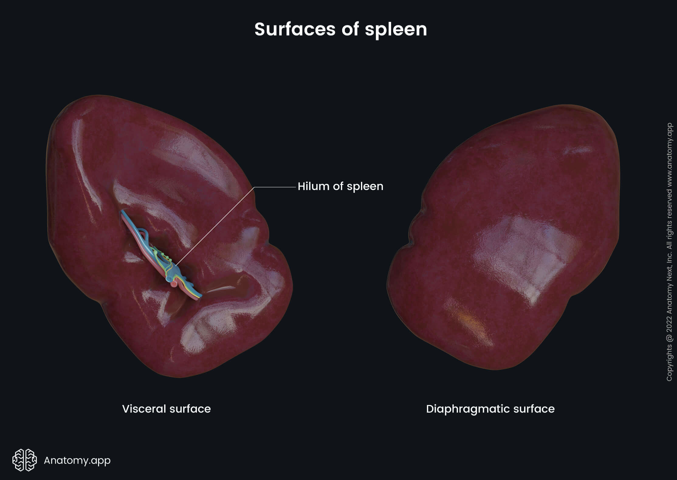
As mentioned previously, the spleen has two surfaces - diaphragmatic and visceral.
Diaphragmatic surface
The diaphragmatic surface is the posteromedial surface that faces the dome of the left hemidiaphragm. It is smooth and slightly convex to adjust the concavity of the diaphragm.
The diaphragmatic surface contacts not only the inferior surface of the diaphragm but also the rib cage. Therefore, it has impressions that are formed by the ribs nine to eleven.
Visceral surface
The visceral surface of the spleen is directed medially. Opposite to the diaphragmatic surface, it is in contact with other internal organs of the abdomen, such as the stomach, colon, pancreas and left kidney.
This surface is concave and irregular, containing several impressions created by the mentioned organs. Also, the central aspect of the visceral surface has the hilum of the spleen.
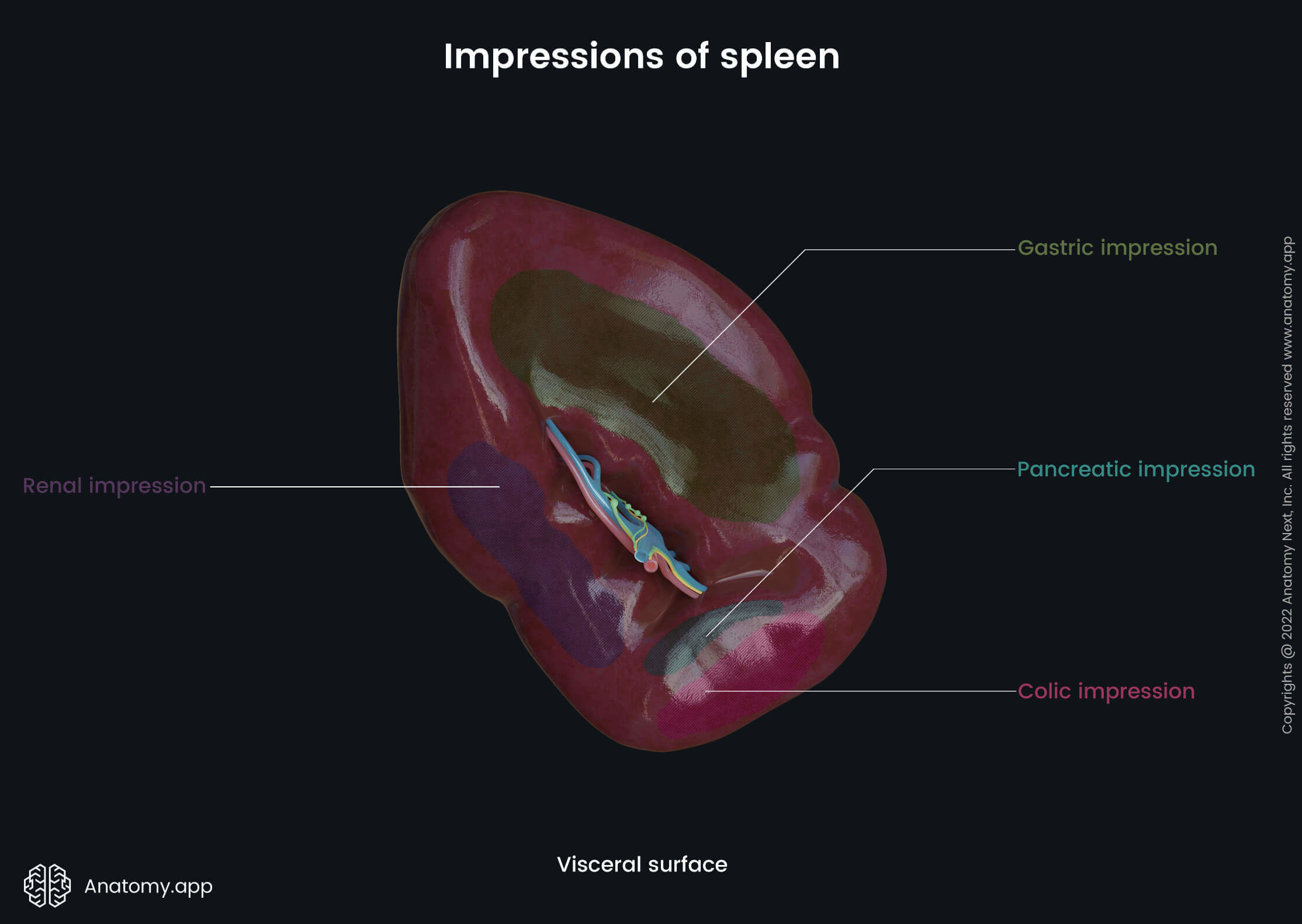
The visceral surface presents with the following impressions:
- Gastric impression - found anteromedially between the superior border of the spleen and the splenic hilum; it is the site where the spleen connects with the fundus of the stomach; the most concave impression;
- Colic impression - found at the inferior aspect of the visceral surface; marks the site where the spleen connects with the left colic (splenic) flexure;
- Pancreatic impression - found medially between the hilum of the spleen and the colic impression; it is located right under the inferior aspect of the hilum of the spleen; marks the site where the tail of the pancreas touches the spleen;
- Renal impression - found posteromedially between the inferior border and hilum of the spleen; corresponds to the site where the left kidney connects with the spleen.
Note: Some authors use words like areas or surfaces instead of impressions, and it is not incorrect. However, sites where the spleen connects with other organs are slightly depressed; therefore, we prefer to use the term impressions.
Spleen borders
Previously it was known that the spleen has two distinct borders - superior and inferior. However, some authors lately state that there is one more - intermediate border. We will review all three of them.
- The superior border is the sharpest. Close to the anterior extremity, it has one or two notches. It is located at the level of the ninth rib. The superior border separates the diaphragmatic surface from the gastric impression of the visceral surface.
- The inferior border is relatively round. It is positioned at the level of the eleventh rib. This border separates the renal impression of the visceral surface from the diaphragmatic surface.
- The intermediate border appears thick and incomplete, extending from the posterior extremity to the hilum of the spleen. The intermediate border is found on the visceral surface of the spleen. It separates the gastric impression from the renal impression.
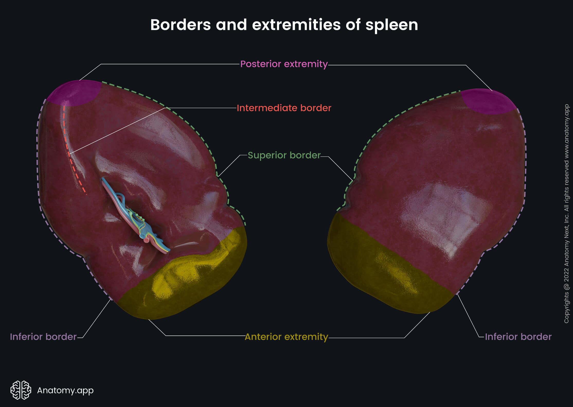
Spleen extremities
Besides the borders, the spleen has two extremities - anterior and posterior. The anterior extremity is the lower pole, and it is also sometimes called the lateral extremity. It is directed downward and forward.
In contrast, the posterior extremity is the upper pole, and it is also known as the medial extremity. It is directed upward, backward and medially. Overall, the anterior extremity is more expanded than the posterior.
Splenic capsule
As mentioned before, the spleen is surrounded by the peritoneum. Underneath the peritoneum, the spleen is encased by a weak outer connective tissue capsule containing dense irregular fibroelastic tissue.
Overall, the splenic capsule has a protective function, and it helps the organ to remain in place. At the same time, the capsule also contains contractile cells called myofibroblasts. Their contractions help to discharge the blood within the spleen into the circulation.
Moreover, the spleen capsule is capable of increasing in size significantly when necessary. Additionally, the connective tissue of the capsule extends into the spleen parenchyma. These septae are called trabeculae, and they subdivide the splenic parenchyma into smaller internal sections.
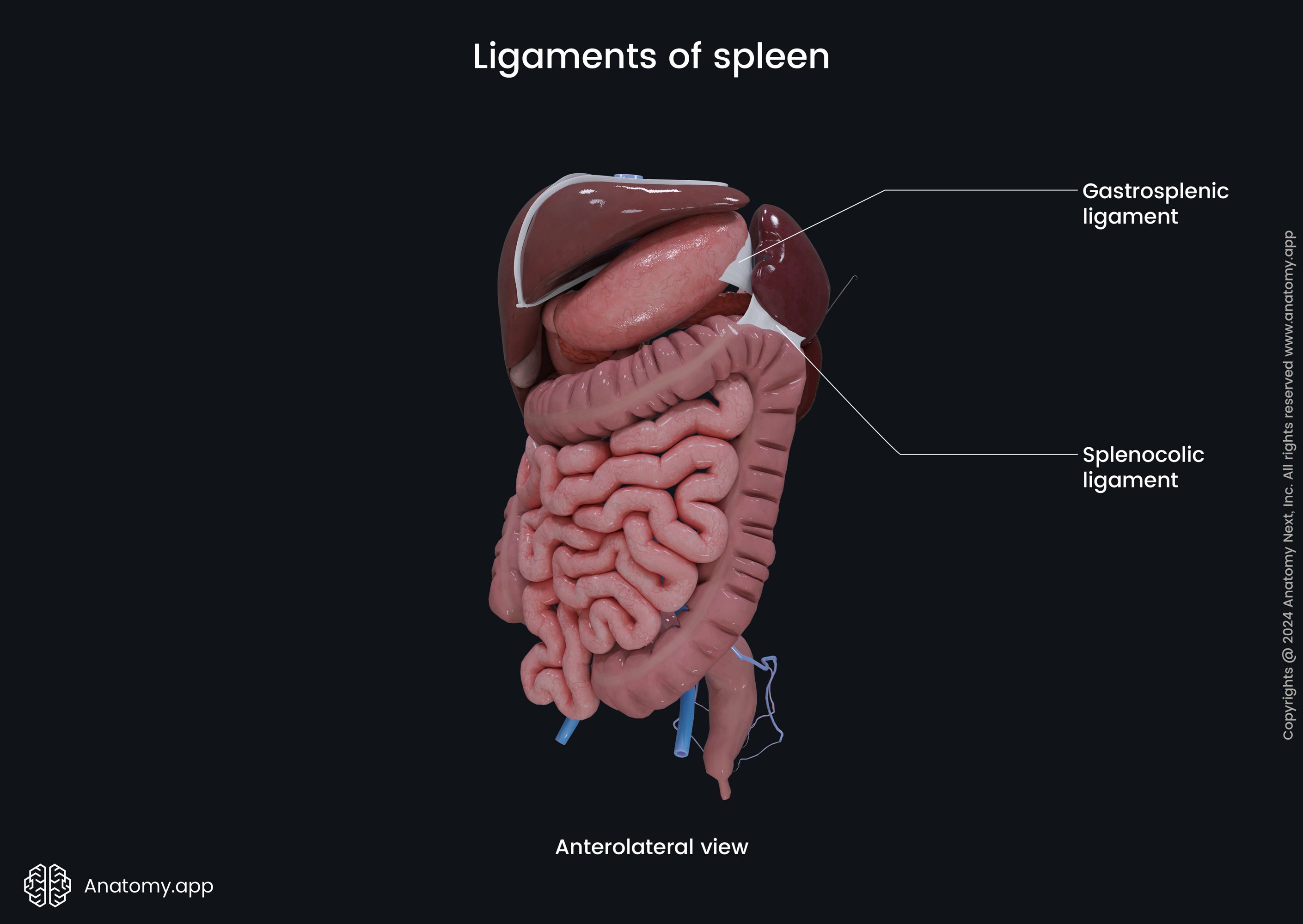
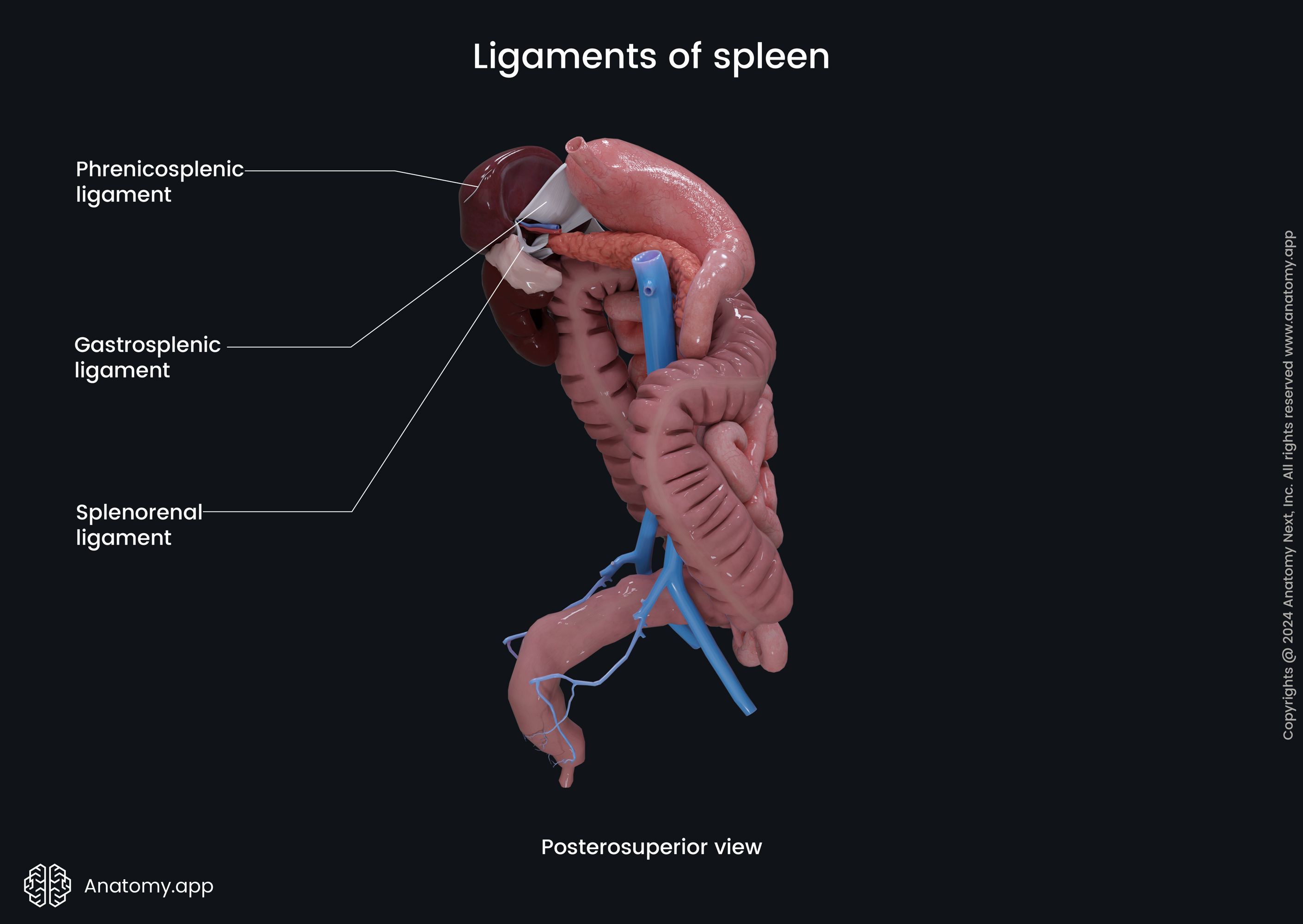
Spleen ligaments
The spleen is fixed in its place by several ligaments, surrounding organs, and intra-abdominal pressure. Overall, the spleen is connected to three main ligaments originating from the surrounding structures and peritoneum. One more ligament is not directly connected to the spleen but plays an essential role in its fixation.
- Gastrosplenic ligament - passes anterior to the splenic hilum, connects the hilum with the greater curvature of the stomach; contains short gastric and the left gastroepiploic (gastroomental) vessels;
- Splenorenal ligament - extends posteriorly to the splenic hilum; connects the splenic hilum to the left kidney; the splenic vessels and also the tail of the pancreas lie within this ligament; it transmits the splenic artery and vein;
- Phrenicosplenic ligament - connects the inferior surface of the diaphragm to the posterior extremity of the spleen;
- Phrenicocolic ligament - connects the left colic flexure with the diaphragm; the superior surface of the ligament supports the anterior extremity of the spleen.
Spleen histology
The spleen is an encapsulated organ of the lymphatic system that filters and immunologically monitors blood. Like other internal organs, the spleen also consists of stroma and parenchyma.
The stroma of the spleen is mainly composed of reticular connective tissue. The parenchyma is called the pulp, and it is subdivided into two functionally and morphologically distinct compartments - white and red pulp. Both are different in their composition, vascular organization and architecture.
Overall, the red pulp is responsible for the filtration of the blood that flows through the spleen. It removes foreign particles and senescent and damaged erythrocytes. Also, it stores platelets, erythrocytes and iron. Moreover, the red pulp is a site for hematopoiesis during the fetal period. In contrast, the white pulp contains lymphocytes and initiates an immune response.
White pulp
The white pulp is the central portion of the spleen. It is mainly formed by the lymphatic tissue that contains white blood cells. The white pulp is located around blood vessels as longitudinal formations and round nodes.
The white pulp of the spleen is composed of three different compartments - periarteriolar lymphoid sheath (PALS), lymphoid follicles and the marginal zone:
- The periarteriolar lymphoid sheath (PALS) is composed of a central artery and surrounding lymphoid tissue. The central artery is a branch of the splenic artery. Lymphoid tissues are organized into two layers - inner and outer. The inner layer is also called the T-zone, as it contains T lymphocytes. The outer zone contains both - B and T lymphocytes. It also contains macrophages and plasma cells.
- The lymphoid follicles are found adjacent to the branches of the central arterioles. They are formed primarily by B lymphocytes and can be subdivided into primary and secondary follicles. The primary follicle is composed mainly of small and immature lymphocytes. The secondary follicles arise from the primary follicles, and they contain mature lymphocytes. The secondary follicles have distinctive germinal centers. They are sites where lymphocytes mature. Besides the B lymphocytes, the germinal centers also house follicular dendritic cells.
- The marginal zone is the peripheral area of the white pulp found at the interface of the red pulp with the lymphoid follicles and PALS. It contains various immune cells such as macrophages, dendritic cells and lymphocytes. Mentioned cells interact and provide an immune response.
Overall, the primary function of the white pulp is immune surveillance.
Note: Some authors consider the marginal zone rather as a separate compartment than a part of the white pulp.
Red pulp
The majority of the spleen is formed by the red pulp. The red pulp consists of the splenic sinusoids and the cords of Billroth (splenic cords). Besides large amounts of sinusoids, the red pulp also contains massive amounts of macrophages.
- The splenic cords or cords of Billroth are cellular aggregations consisting of macrophages, lymphocytes, and plasmocytes. These cords appear as stripes, and they are supported by the reticular connective tissue.
- The splenic sinusoids are positioned between the splenic cords. They are blood-filled cavities, giving the red pulp its red color. As blood flows through the sinusoids, the macrophages from the splenic cords eliminate the foreign antigens and old and damaged red blood cells.
Overall, the main function of the red pulp is blood filtration. It serves as a blood filter that eliminates various antigens before they further enter the systemic circulation.
Neurovascular supply of spleen
As mentioned, the spleen is a highly vascular organ with a substantial vascular network. The innervation of this organ is provided by the sympathetic and parasympathetic nervous systems of the autonomic nervous system.
Arterial blood supply
The spleen receives arterial blood supply from the splenic artery, which is a branch of the celiac trunk. The celiac trunk itself arises from the abdominal aorta. The splenic artery reaches the spleen running within the splenorenal ligament along the superior aspect of the pancreas.
As the splenic artery reaches the spleen, it divides into five branches that provide arterial blood supply to different regions of the organ. All of these branches do not anastomose with each other, and it results in vascular segmentation of the spleen.
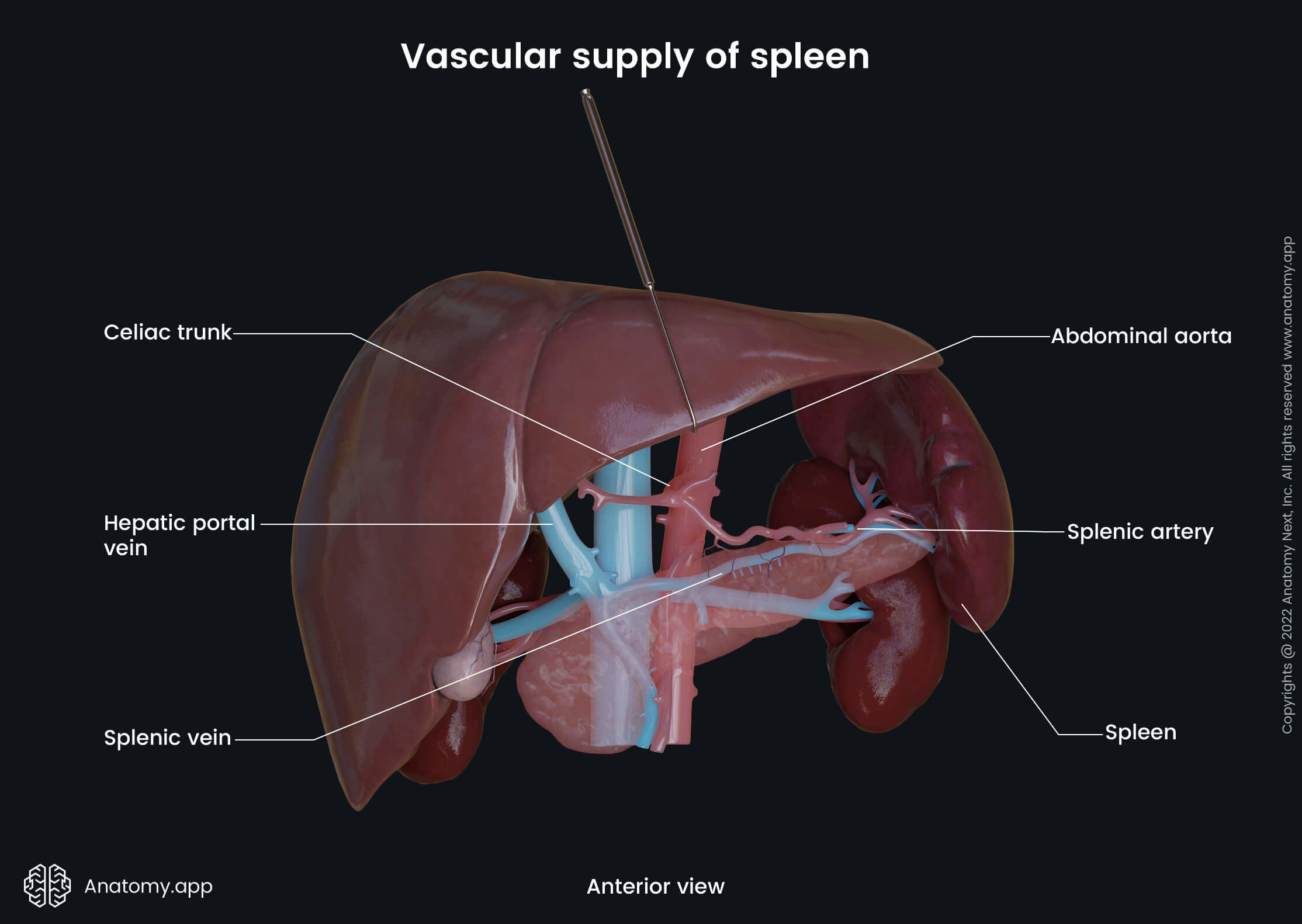
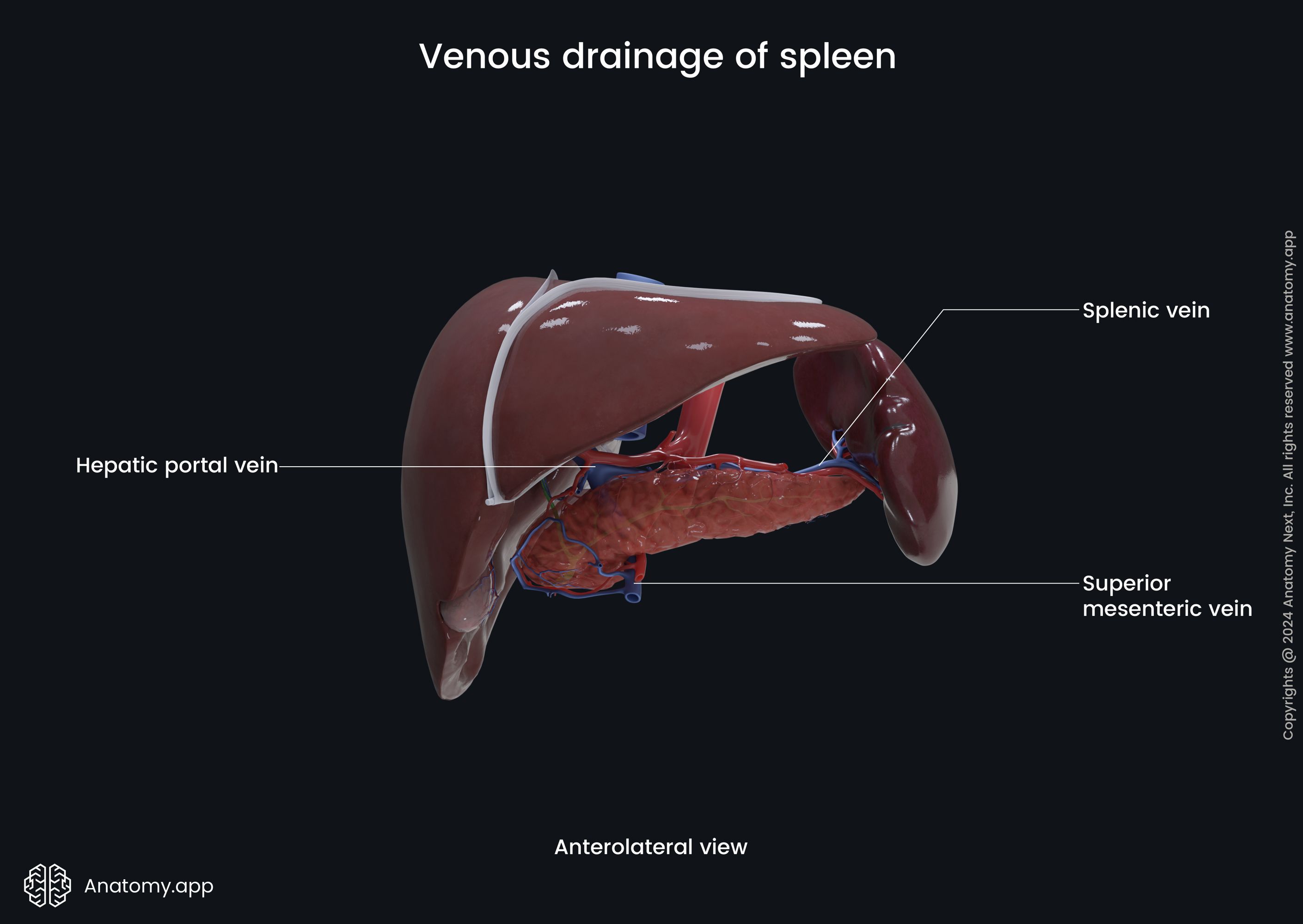
Venous drainage
The venous drainage of the spleen is provided by the splenic vein. It travels within the splenorenal ligament below the splenic artery. The splenic vein carries collected venous blood to the hepatic portal vein, which is formed at the level of the neck of the pancreas due to the merging of the splenic vein and the superior mesenteric vein.
Lymphatic drainage
Lymphatic drainage is provided by the efferent lymphatic vessels that travel alongside the splenic vein and splenic artery. They drain into the pancreaticosplenic lymph nodes found on the superior aspect of the pancreas. Further, lymph is carried to the celiac lymph nodes.
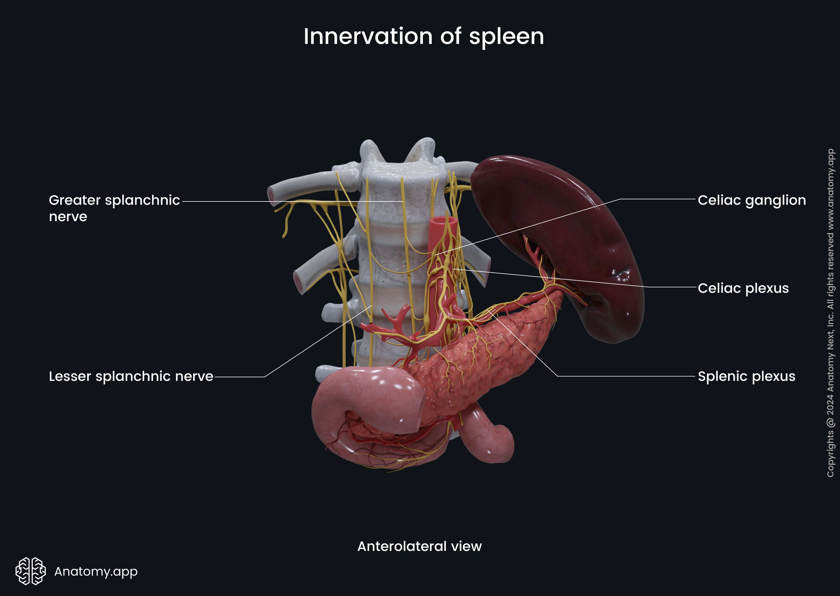
Innervation
The spleen receives sympathetic and parasympathetic innervation via the nerves coming from the celiac plexus. Sympathetic innervation is provided by the greater and lesser splanchnic nerves of the sympathetic trunk, while parasympathetic innervation comes from the vagus nerve (CN X).
Functions of spleen
The spleen has several functions. While it is an essential organ, it is not a vital one. A person can live everyday life without a spleen. In that case, other internal organs will adapt to its absence and provide about the same functions. As mentioned, the spleen is composed of white and red pulp, each providing a different range of functions.
- During the fetal period, the spleen is partially responsible for hemoglobin synthesis and hematopoiesis. It usually actively happens from the 10th to the 25th week of gestation. Later, the bone marrow takes over the process.
- The white pulp primary is involved in the production and maturation of white blood cells, particularly B and T lymphocytes. Therefore, white pulp also is responsible for the production of antibodies. Antigens are presented by antigen-presenting cells within the lymphoid follicles of the white pulp. In turn, this process initiates the activation of T and B lymphocytes, leading to the synthesis of antibodies.
- In contrast, the red pulp is a structure that provides blood filtration. The red pulp not only removes senescent and damaged red blood cells but also eliminates various antigens, as the red pulp also contains white blood cells and macrophages that provide phagocytosis and destroy foreign microorganisms.
- Also, it is involved in iron metabolism, as splenic macrophages are specialized to recycle iron during blood filtration from the old and damaged red blood cells. This process is essential for synthesizing hemoglobin. Macrophages will then store ingested iron or export it into the bloodstream via ferritin.
- Moreover, the spleen acts as a reservoir for blood. The red pulp serves as a storage site for white blood cells and platelets. The spleen can contract its capsule and trabeculae to increase systemic blood supply due to sympathetic stimulation during hemorrhage or shock. The spleen stores approximately 25 - 30% of red blood cells and 25 - 30% of platelets.
- And finally, the spleen can also play a role in hematopoiesis if required. It can provide extramedullary hematopoiesis during various pathologic conditions (such as beta-thalassemia major). And this will help the bone marrow to compensate for the hemolysis that is occurring.
Anatomy.app
Contact information
- For questions regarding business inquiries. Please contact:
- info@anatomy.app