- Anatomical terminology
- Skeletal system
- Joints
- Muscles
- Head muscles
- Neck muscles
- Muscles of upper limb
- Thoracic muscles
- Muscles of back
- Muscles of lower limb
- Heart
- Blood vessels
- Lymphatic system
- Nervous system
- Respiratory system
- Digestive system
- Urinary system
- Female reproductive system
- Male reproductive system
- Endocrine glands
-
Eye
- Accessory structures of eye
- Ear
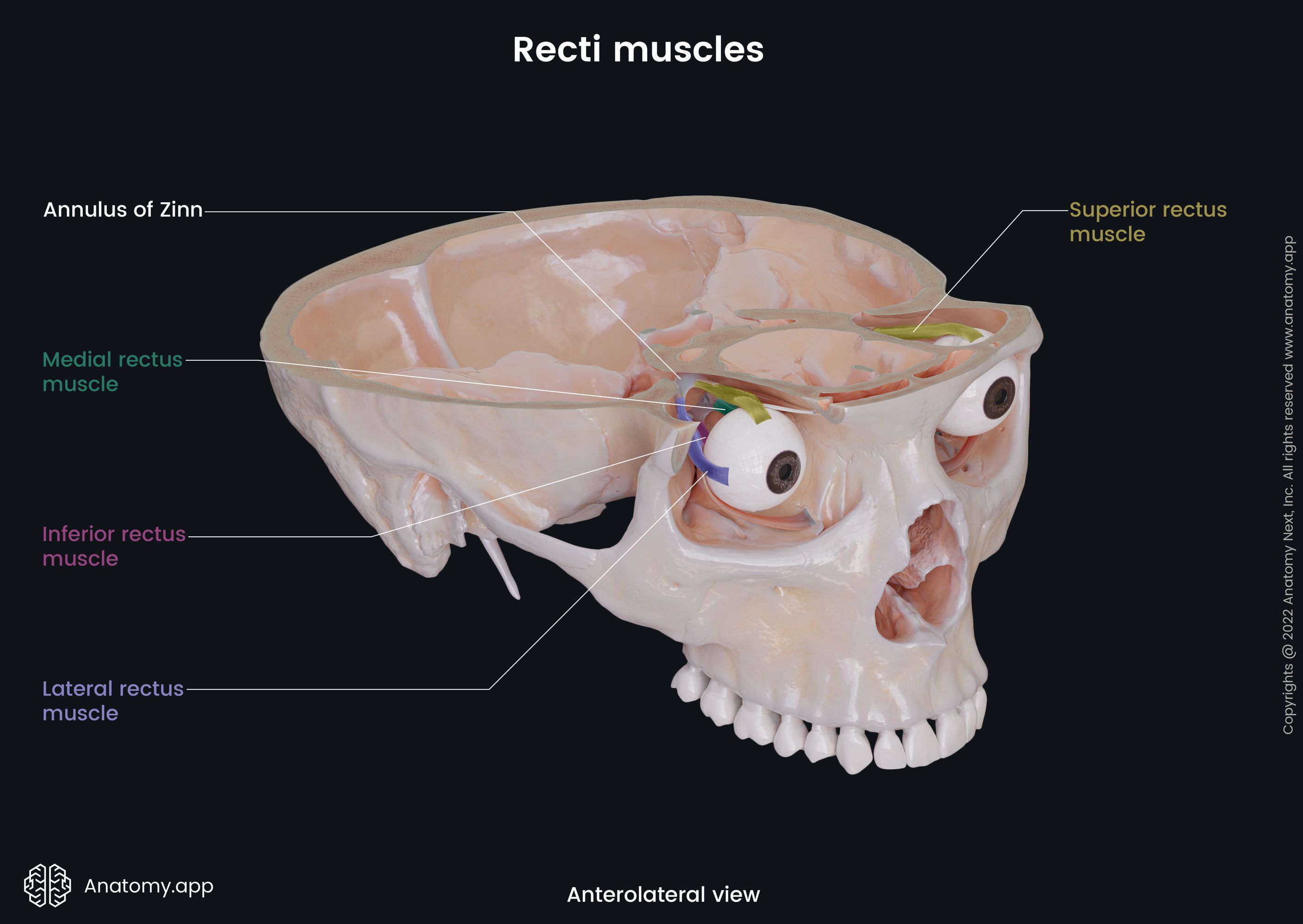
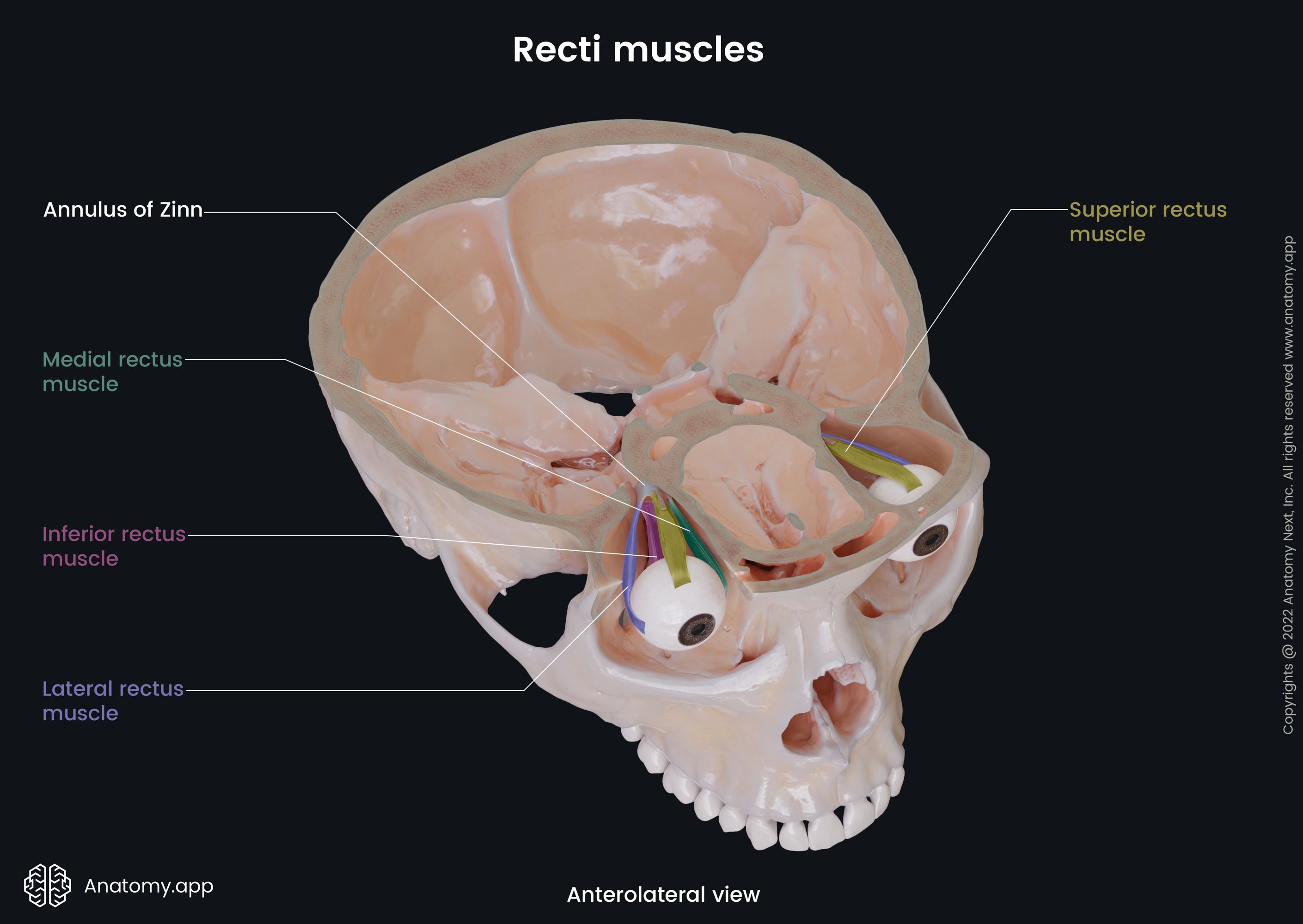
Superior rectus
The superior rectus (Latin: musculus rectus superior), also known as the superior rectus extraocular muscle, is one of the six extraocular muscles that are in control of eye movements. Actions of the superior rectus result in moving the visual gaze up and in.
Origin
The superior rectus originates from the upper part of the common tendinous ring, above and lateral to the optic canal. Some fibres of the superior rectus also arise from the dural sheath of the optic nerve (CN II).
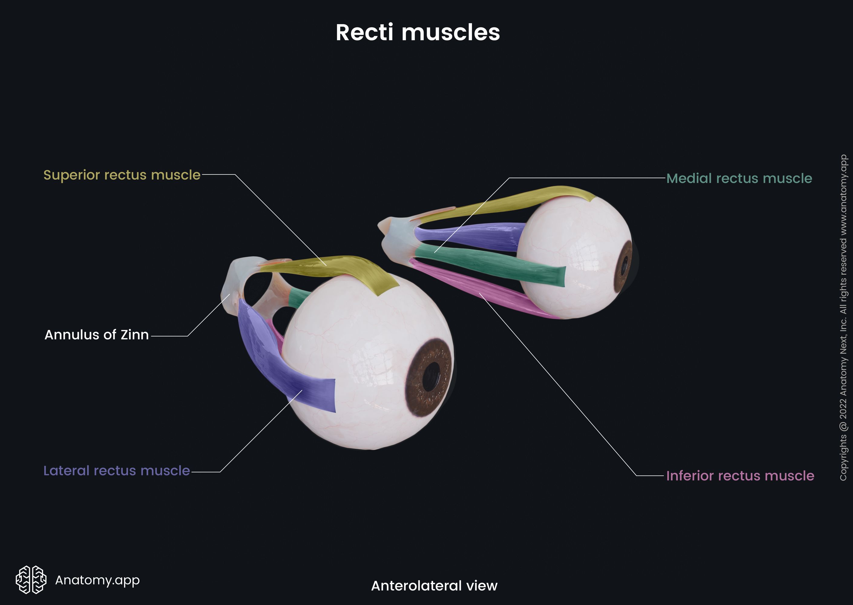
Insertion
The superior rectus inserts into the upper part of the sclera, approximately 0.3 inches (8 mm) from the corneal limbus.
Action
The main actions provided by the superior rectus are elevation and adduction of the eyeball, and medial rotation of the eyeball. To obtain the upward movement, the muscle must function in synergy with the inferior oblique.
Innervation
The superior rectus is innervated by the superior division of the oculomotor nerve (CN III).
Blood supply
The arterial blood supply to the superior rectus is provided by the ophthalmic artery and indirectly from its supraorbital branch.
Anatomy.app
Contact information
- For questions regarding business inquiries. Please contact:
- info@anatomy.app