- Anatomical terminology
- Skeletal system
- Joints
- Muscles
- Head muscles
- Neck muscles
- Muscles of upper limb
- Thoracic muscles
- Muscles of back
- Muscles of lower limb
- Heart
- Blood vessels
- Lymphatic system
- Nervous system
- Respiratory system
- Digestive system
- Urinary system
- Female reproductive system
- Male reproductive system
- Endocrine glands
-
Eye
- Accessory structures of eye
- Ear
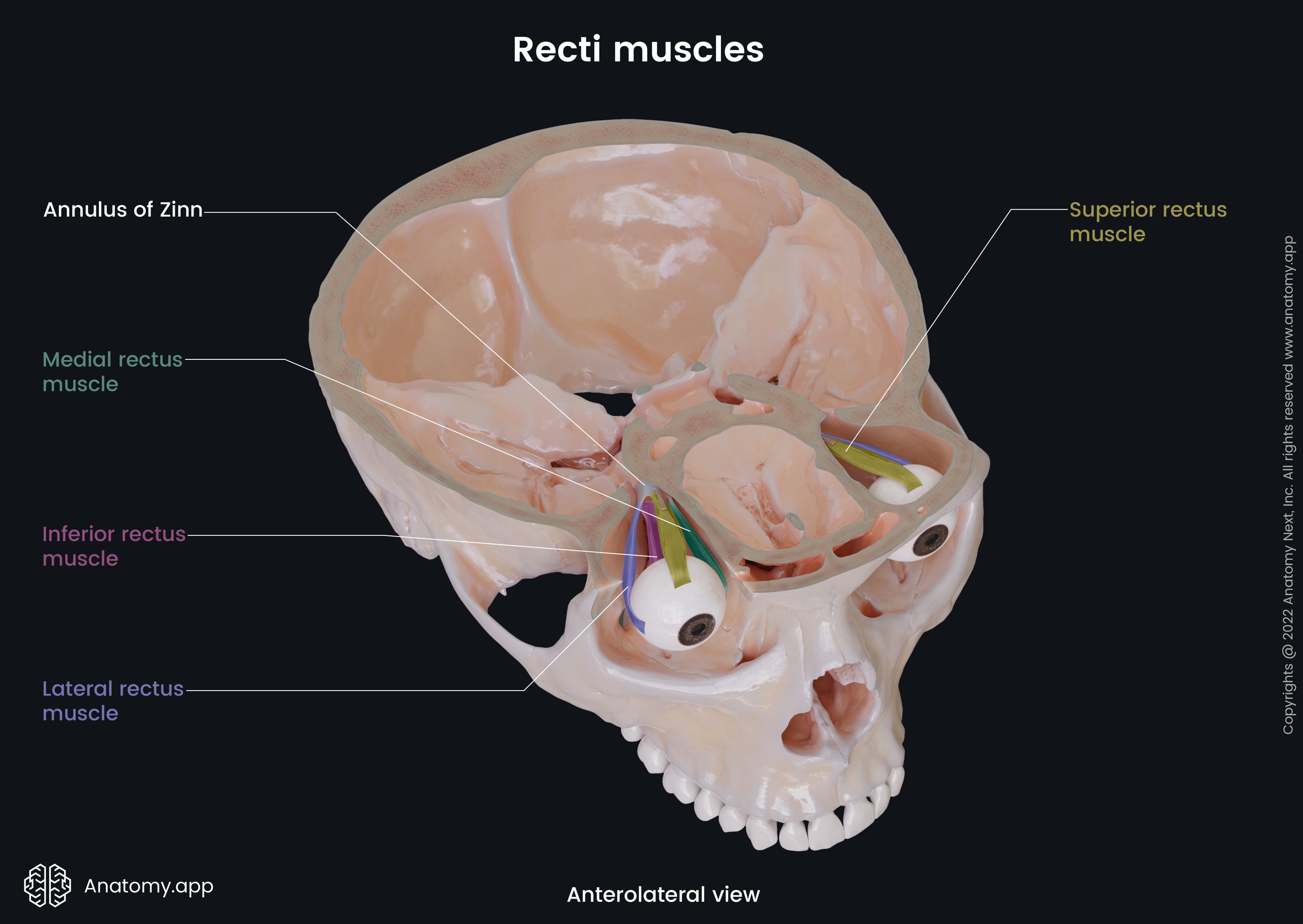
Medial rectus
The medial rectus (Latin: musculus rectus medialis), also called the medial rectus extraocular muscle, is one of the six extraocular muscles that are in control of eye movements. The medial rectus is responsible for turning the visual gaze medially.
Origin
The medial rectus originates from the medial part of the common tendinous ring, and also from the dural sheath of the optic nerve (CN II). The muscle passes horizontally forwards along the medial wall of the orbit, below the superior oblique muscle.
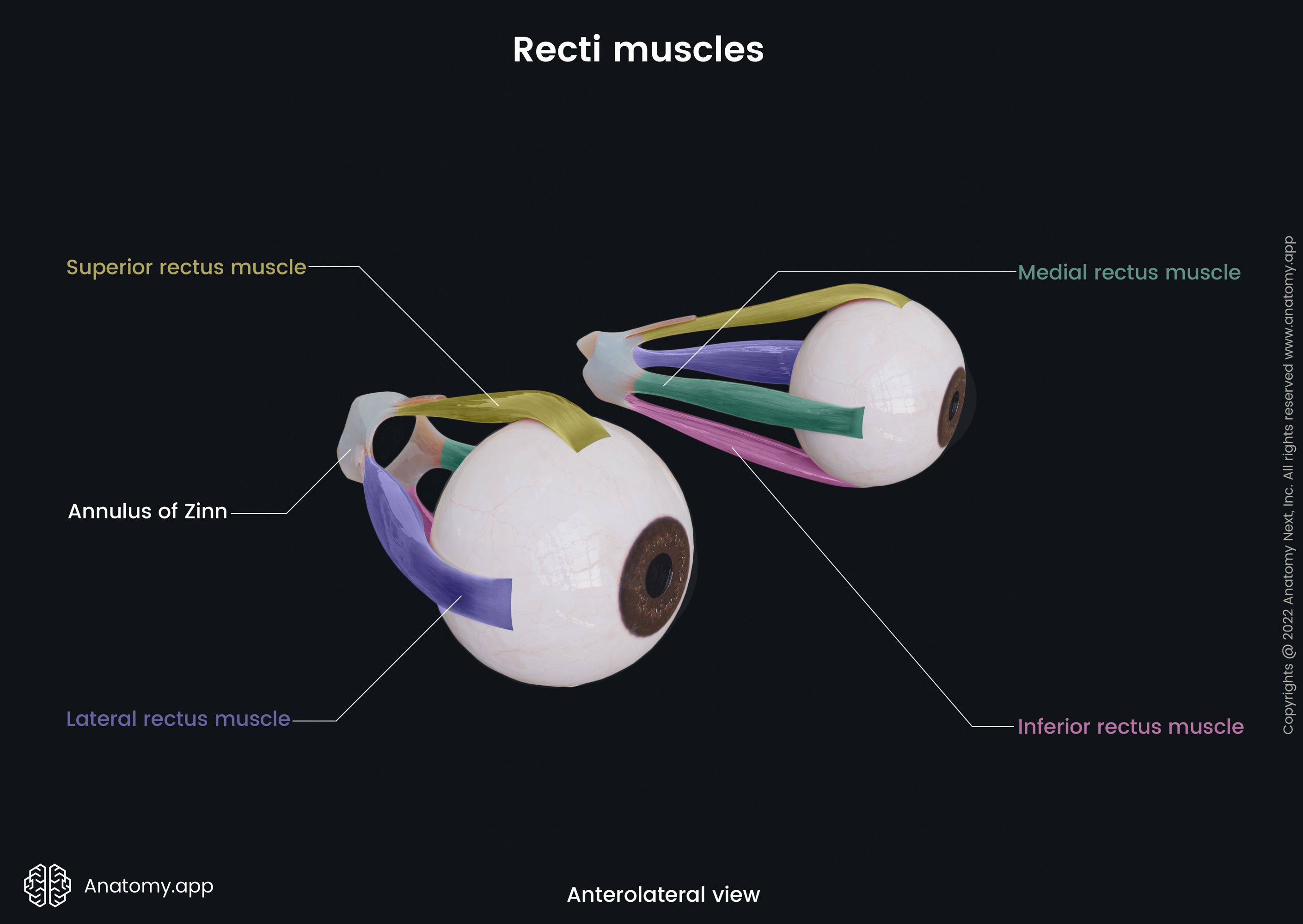
Insertion
The medial rectus inserts into the medial surface of the sclera, approximately 0.2 inches (5.5 mm) from the limbus and slightly anterior to the other recti muscles.
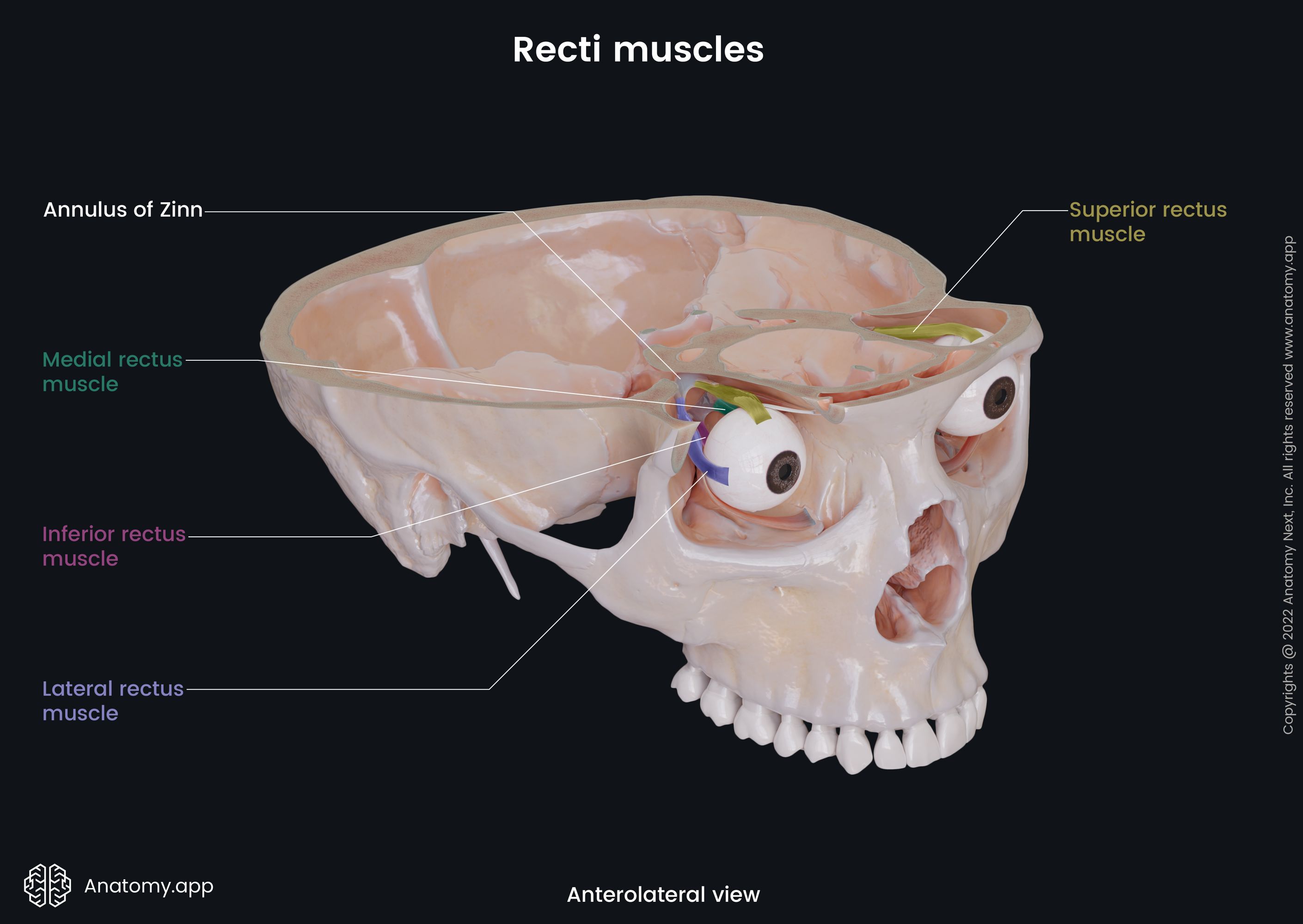
Action
Contractions of the medial rectus turn the eyeball medially. The right and left medial recti acting together are responsible for convergence of the eyes - moving the eyes closer together and turning inward to look at a close object.
Innervation
The medial rectus is innervated by the oculomotor nerve (CN III).
Blood supply
The medial rectus receives arterial blood supply mainly from the ophthalmic artery.
Anatomy.app
Contact information
- For questions regarding business inquiries. Please contact:
- info@anatomy.app