- Anatomical terminology
- Skeletal system
- Joints
- Muscles
- Heart
- Blood vessels
- Blood vessels of systemic circulation
- Aorta
- Blood vessels of head and neck
- Blood vessels of upper limb
- Blood vessels of thorax
- Blood vessels of abdomen
- Arteries of abdomen
- Veins of abdomen
- Blood vessels of pelvis and lower limb
- Blood vessels of systemic circulation
- Lymphatic system
- Nervous system
- Respiratory system
- Digestive system
- Urinary system
- Female reproductive system
- Male reproductive system
- Endocrine glands
- Eye
- Ear
Abdominal aorta
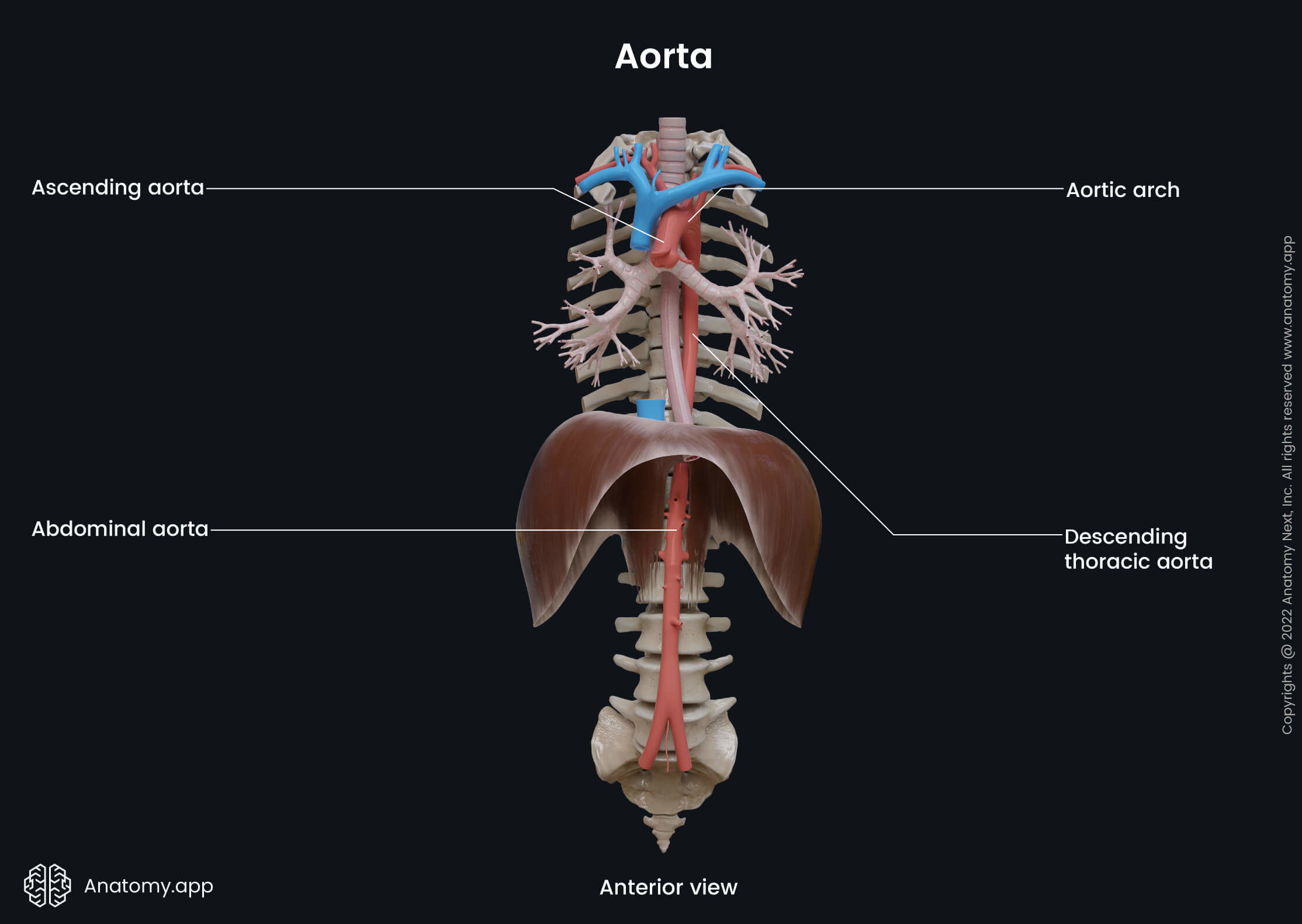
The abdominal aorta (Latin: aorta abdominalis) is the abdominal part of the descending aorta and the largest artery in the abdomen. It is the continuation of the thoracic aorta (thoracic part of the descending aorta) after it enters the abdomen via the aortic hiatus of the diaphragm. The abdominal aorta terminates by dividing into two vessels - the right and left common iliac arteries.

Course of abdominal aorta
The abdominal aorta begins at the aortic hiatus of the diaphragm at the level of the lower part of the 12th thoracic vertebra (T12). The aorta descends along the posterior wall of the abdomen, traveling down the midline along the anterior surface of the bodies of the lumbar vertebrae L1 to L4. The abdominal aorta ends on the left of the midline at the lower level of the vertebra L4, where it divides into the right and left common iliac arteries.
Relations of abdominal aorta
Along its course through the posterior abdominal region, the anterior surface of the abdominal aorta is covered by the prevertebral plexus - a network of nerves and ganglia. The aorta is also related to several other structures in the abdomen:
- Located anterior to the abdominal aorta is the pancreas, as well as the splenic vein, left renal vein, and the inferior part of the duodenum;
- Posteriorly, the abdominal aorta is crossed by several left lumbar veins as they pass to the inferior vena cava;
- Situated on its right side are the cisterna chyli, thoracic duct, azygos vein, right crus of the diaphragm, and the inferior vena cava;
- Located on the left side of the abdominal aorta is the left crus of the diaphragm.
Branches of abdominal aorta
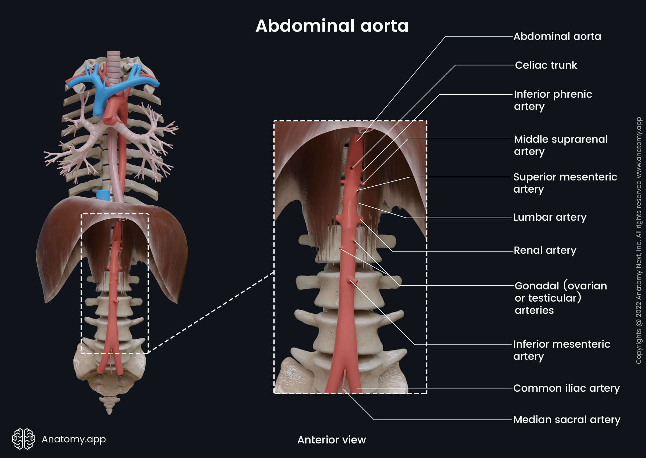
The abdominal aorta gives off many essential branches, mainly to supply the abdominal region with oxygenated blood. These branches can be categorized into three groups:
- Posterior branches - supplying the diaphragm or body wall;
- Visceral branches - supplying internal organs;
- Terminal branches - providing mainly the pelvis and lower limb.

The posterior branches of the abdominal aorta supply the diaphragm or the body wall and include the following arteries:
There are paired and unpaired visceral branches arising from the abdominal aorta. Three unpaired visceral branches originate from the anterior surface of the aorta. They are also known as the anterior branches of the abdominal aorta and include the following arteries:
The paired visceral branches arising on each side of the abdominal aorta include the following:
- Middle suprarenal arteries
- Renal arteries
- Testicular (in males) or ovarian arteries (in females)
As mentioned previously, the terminal branches of the abdominal aorta are the two common iliac arteries (right and left) arising from its bifurcation. The bifurcation usually occurs at the level of the fourth lumbar vertebra (L4).