Anatomy.app Content News in July
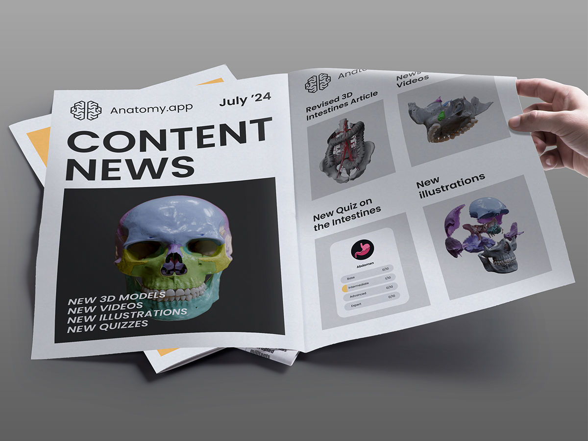
Our dedicated team of professionals has been tirelessly working in the previous month to revise, update, and expand our content to provide our subscribers with the best learning experience possible. So, we are excited to share the latest updates and additions to Anatomy.app in July! Browse the new 3D models, videos, illustrations, and quizzes!
Here's a detailed look at what’s new:
1. 3D Anatomy: Revised Intestines Article
We have thoroughly revised our 3D Intestines article to include more detailed 3D models and comprehensive textual descriptions. This update guarantees an even deeper understanding of the anatomy of the intestines, helping our subscribers better visualize this subject and learn it more effectively.
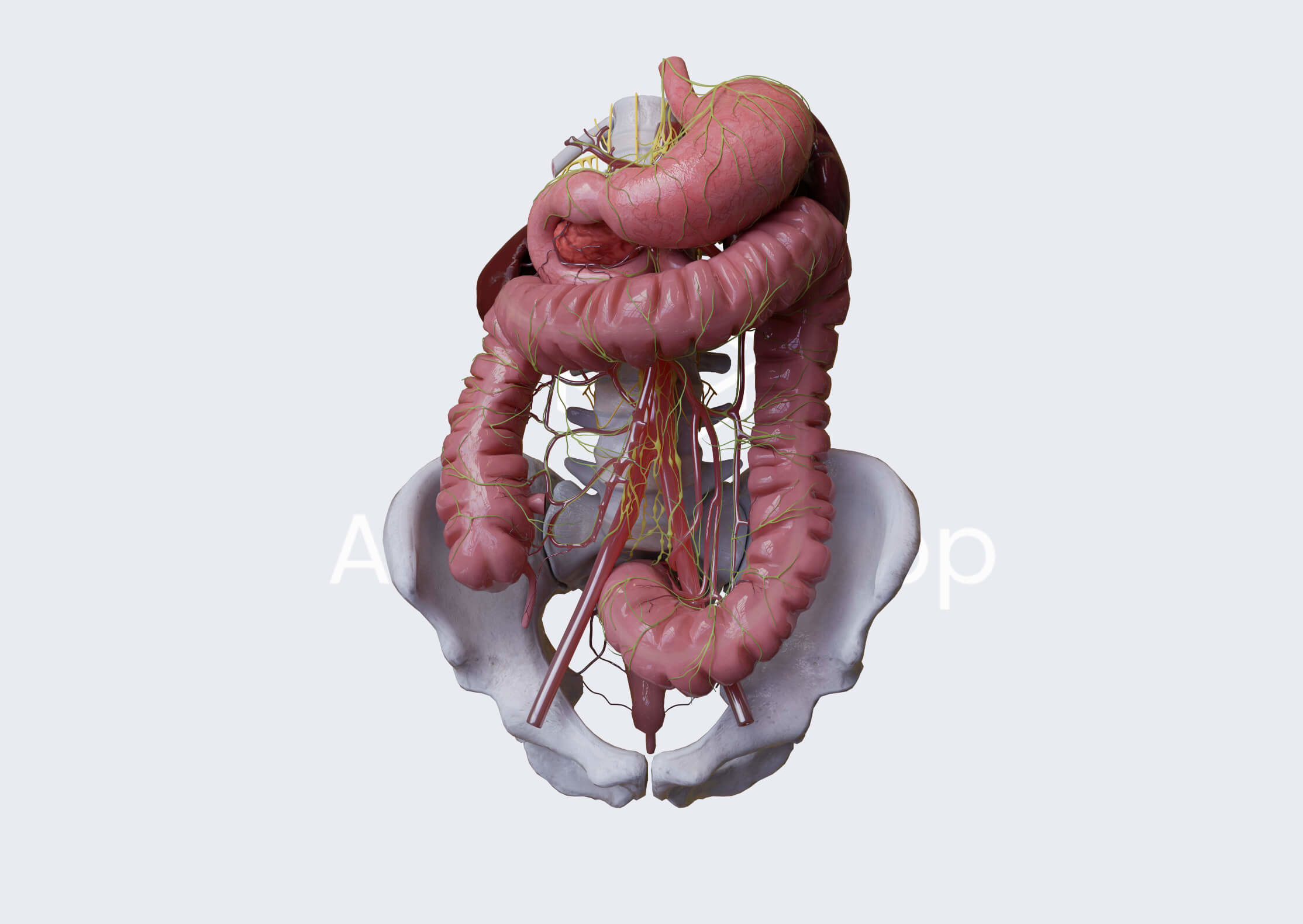
We have added new slides to the article to enrich the learning experience. Now, the 3D Intestines article features 29 slides. All previous 3D models have been meticulously updated, and new models have been added for greater detail and accuracy. Also, all textual information has been thoroughly revised to provide the most comprehensive understanding.
The revised 3D article covers topics such as an overview of the intestines and their parts. It describes the small intestine, including all three parts - duodenum, jejunum, and ileum. Also, it reviews the large intestine, each of its parts, and its characteristic features. In addition, this article also covers arterial blood supply, venous drainage, and innervation of the intestines. Moreover, it reviews various anastomotic networks found in the intestines.
Explore our newly updated 3D Intestines article and elevate your anatomical knowledge to the next level: https://anatomy.app/article/intestines
2. Media Library: a Bunch of New Skull Videos
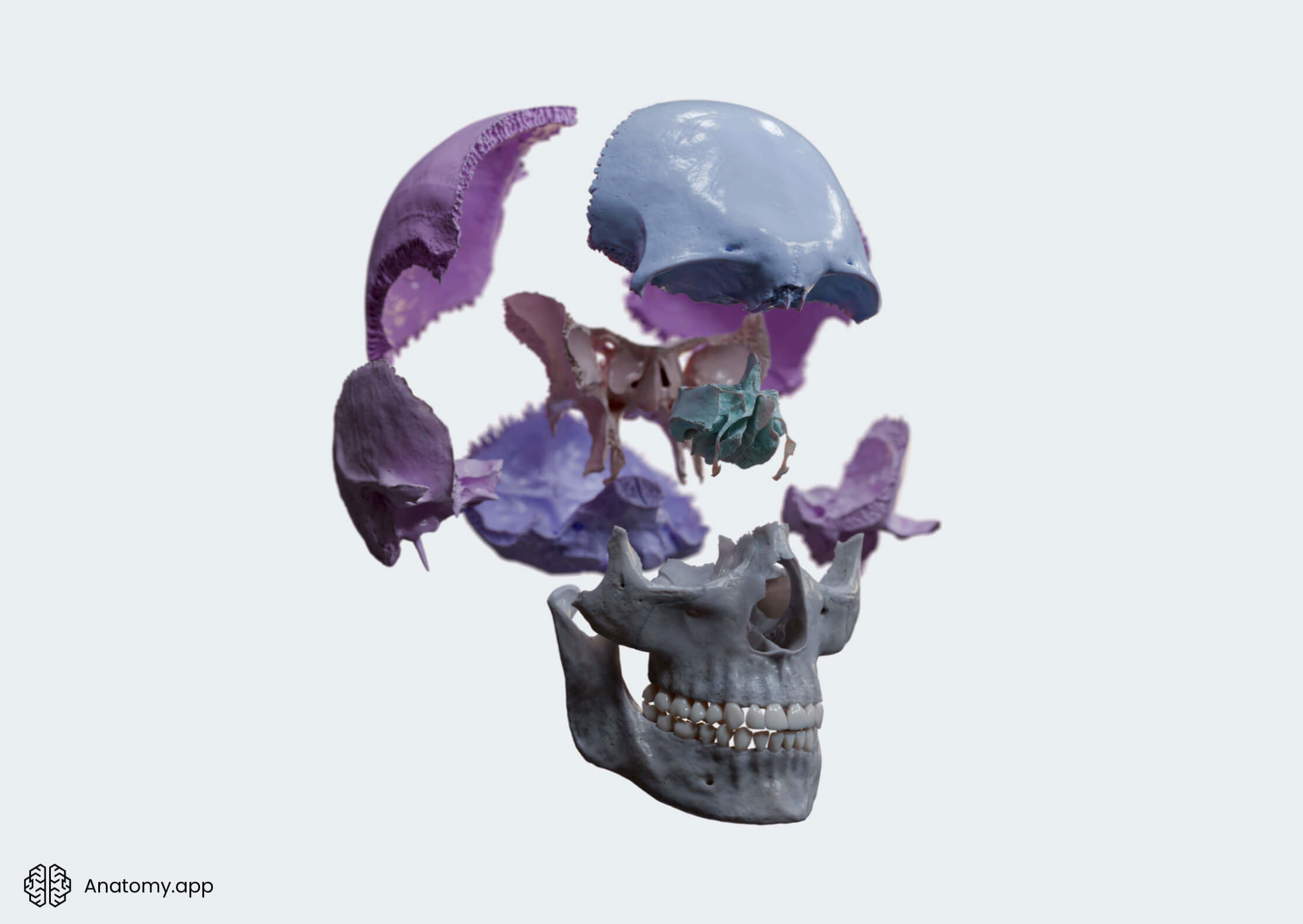
We have expanded our Media Library by adding a bunch of new educational videos that cover various bones of the skull. These videos are designed to help you grasp complex anatomical topics with ease. From now on, no more struggling with learning the bones of the skull. We have got your back!
When browsing through the Media Library, search for the following videos:
- Skull: a 360-degree view of the skull and its bones.
- Skull bones (overview): another 360-degree view of the skull. Bones in this video have been color-coded.
- Skull bones: a comprehensive overview of the bones of the skull. For a deeper and better understanding, skull bones have been color-coded and labeled.
- Skull (360 view): another engaging 360-view of the skull.
- Exploding skull: an interesting video that shows the skull in an exploded view.
- Neurocranium (overview): a 360-degree overview of the neurocranium.
- Viscerocranium (overview): two 360-degree view videos of the viscerocranium.
- Bones of the neurocranium: an explanatory video on the bones forming the neurocranium. All bones of the neurocranium have been color-coded.
- Temporal bone (overview): a 360-degree overview of the temporal bone.
- Palatine bone (parts and landmarks): a detailed look at the palatine bone, its location and position, as well as its parts and key landmarks.
- Zygomatic bone: a 360-degree overview of the zygomatic bone.
- Zygomatic bone (landmarks): a detailed video on the zygomatic bone, its location, processes, and landmarks.
- Inferior nasal concha (overview): a 360-degree view of the inferior nasal concha. This video offers a rare chance to see this bone isolated.
- Inferior nasal concha: another 360-degree overview of the inferior nasal concha. This video helps to understand the location and position of the bone.
- Hyoid bone (overview): a comprehensive 360-degree overview of the hyoid bone.
- Hyoid bone (parts): a breakdown of the parts of the hyoid bone. For a better understanding, the parts of the hyoid bone have been color-coded.
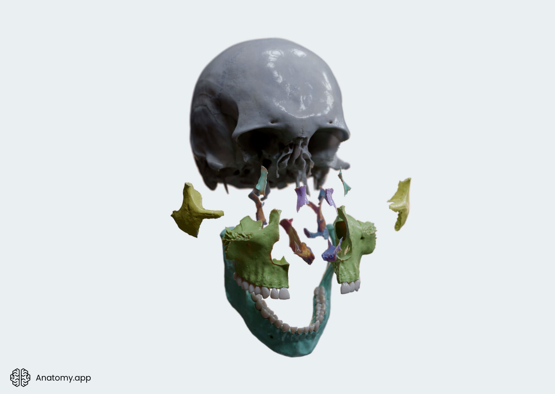
Head to our Media Library, use the filter option, choose the media type (Videos) and the region or organ system (Head and Neck OR Skeletal system), and excel in your anatomy classes.
Watch the new videos now: https://anatomy.app/media?categoryType=regions
3. Encyclopedia: Plenty of New Illustrations
Those large sheets of text without proper illustrations in anatomy are just pieces of paragraphs written in a foreign language, right? That's why we keep updating our online medical library Encyclopedia with new illustrations to aid the understanding of various anatomical structures. This month, you can find a bunch of fresh illustrations in our articles included in the skeletal, muscular, and digestive systems.
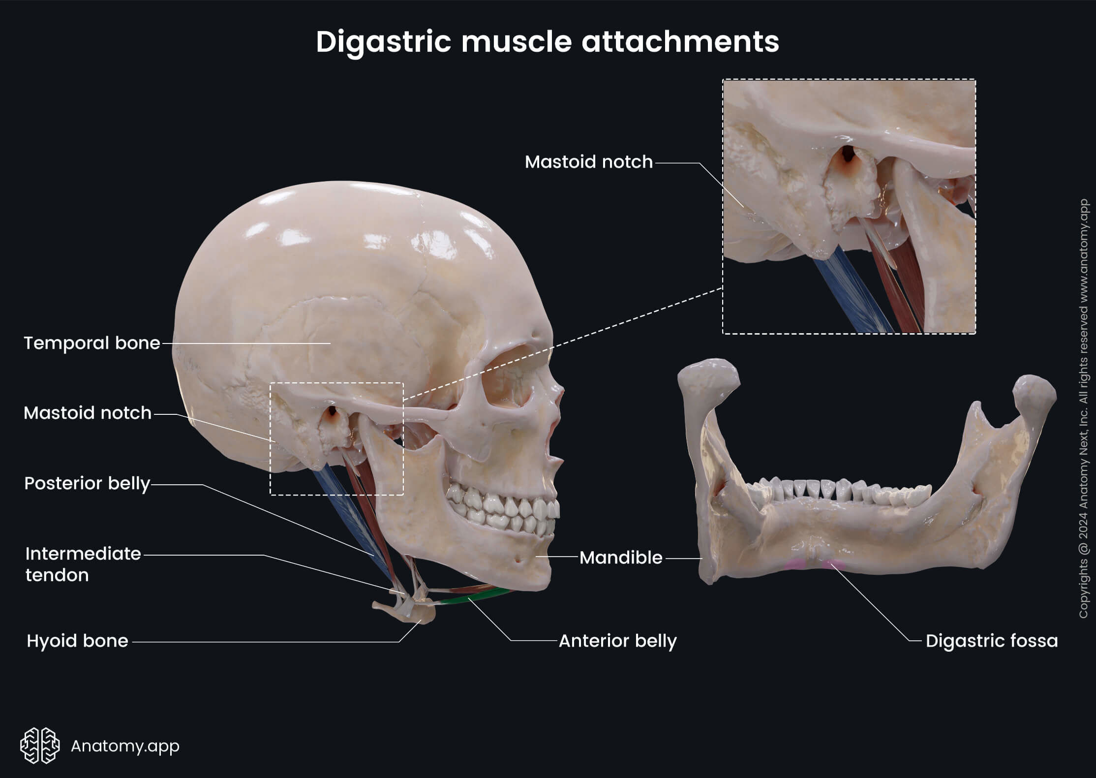
Here is a breakdown of exactly what's been added:
- Frontal bone: a set of detailed illustrations on the parts and landmarks of the frontal bone.
- Parietal bone: several illustrations covering the angles, borders, and landmarks of the parietal bone.
- Stylohyoid: a comprehensive illustration of the stylohyoid and its attachment sites.
- Digastric: an overview of the digastric muscle and its attachment sites.
- Digestive system: a set of illustrations covering the accessory organs of the digestive system.
- Stomach: explanatory illustrations on the pylorus, ligaments, omenta, arterial blood supply, and venous drainage of the stomach.
- Small intestine: a bunch of illustrations covering the parts of the intestines, parts of the small intestine, arterial blood supply, venous drainage, and innervation of the small intestine.
- Duodenum: three illustrations on the arterial blood supply and venous drainage of the duodenum.
- Jejunum and ileum: comprehensive illustrations showing the position and location of the jejunum and ileum, as well as their venous drainage.
- Large intestine: detailed illustrations on the position and location of the large intestine, its parts, arterial blood supply, venous drainage, and innervation.
- Cecum and vermiform appendix: several illustrations covering the neurovascular supply of the cecum and vermiform appendix.
- Colon: a set of illustrations on the parts of the colon, their arterial blood supply and venous drainage.
- Rectum: explanatory illustration on the venous drainage of the rectum.
4. Quizzes: New Quiz on the Intestines
We’ve made a new quiz on the Intestines to match the revised 3D Intestines article. We love quizzes, as they are a fun way on how to test knowledge and enhance learning. Our Intestines quiz is designed in a way that challenges our users, but at the same time, it helps reinforce the understanding of the topic.
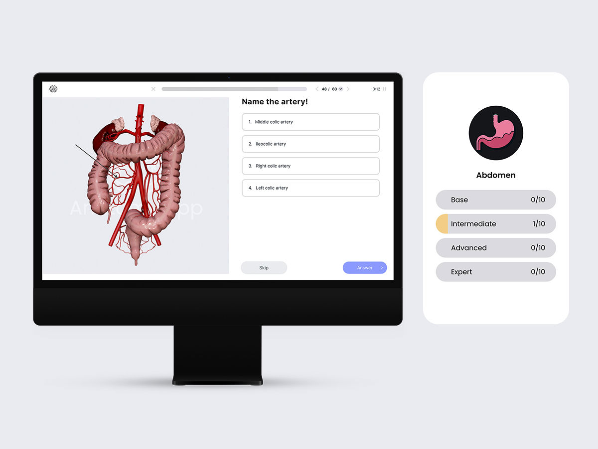
Our Intestines quiz has four difficulty levels, each containing a different number of questions. The base level features 23 questions, the intermediate level has 36 questions, the advanced level includes 52 questions, and the most challenging level - expert - has 60 questions.
Questions in our newly updated Intestines quiz come in various formats to keep our subscribers engaged, entertained, and challenged. Therefore, our Intestines quiz includes different types of questions, such as those with illustrations and without, single-choice, and multiple-choice. This variety ensures a comprehensive assessment of the understanding of intestinal anatomy.
Test your knowledge now: https://anatomy.app/quizzes/topics
Final Note
Whether you are a medical student, professional or an anatomy enthusiast, we hope all these updates will significantly enhance the learning experience on Anatomy.app. With new content updates, we are committed to supporting your journey in understanding human anatomy. Stay tuned for more updates and news as we continue to expand and improve Anatomy.app! Happy exploring and learning!
Best, the Anatomy.app team!🧠