- Anatomical terminology
- Skeletal system
- Joints
- Muscles
- Head muscles
-
Neck muscles
- Superficial neck muscles
- Scalene muscles
- Suprahyoid muscles
- Infrahyoid muscles
- Prevertebral muscles
- Suboccipital muscles
- Muscles of upper limb
- Thoracic muscles
- Muscles of back
- Muscles of lower limb
- Heart
- Blood vessels
- Lymphatic system
- Nervous system
- Respiratory system
- Digestive system
- Urinary system
- Female reproductive system
- Male reproductive system
- Endocrine glands
- Eye
- Ear
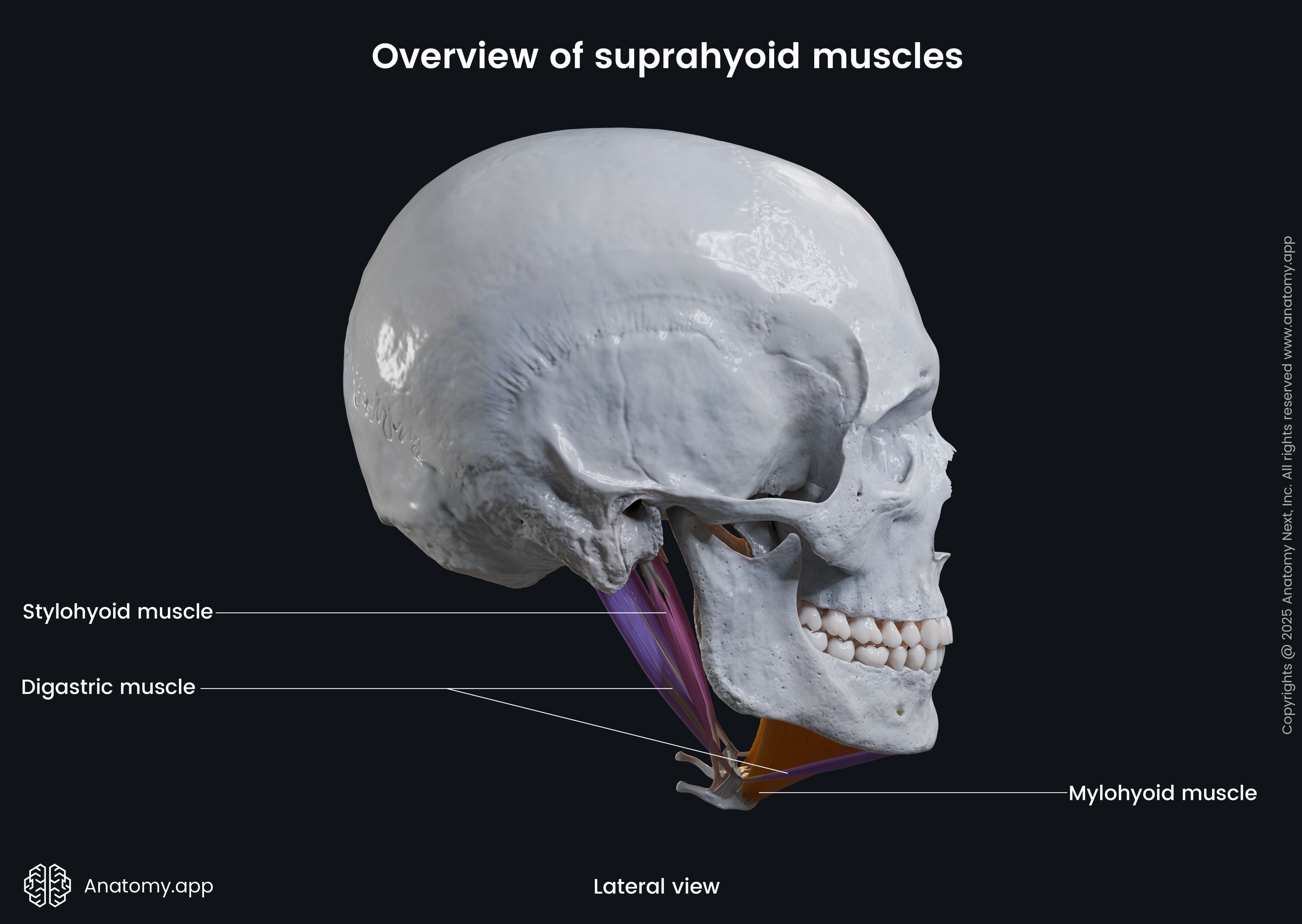
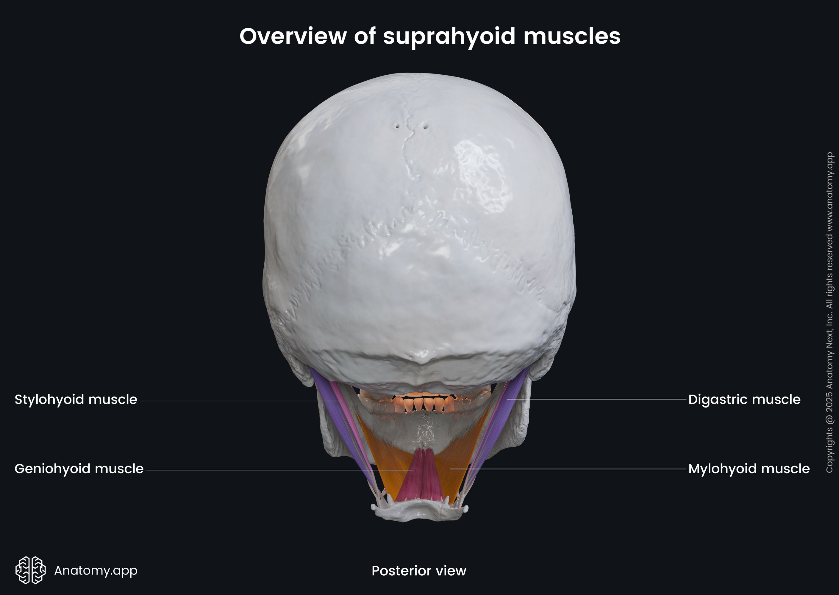
Suprahyoid muscles
The suprahyoid muscles are a group of four paired muscles that are located in the anterior compartment of the neck. As the name suggests, they lie above the hyoid bone. These muscles attach to the superior aspect of the hyoid bone and connect it to the mandible and temporal bone. Additionally, some of these muscles contribute to forming the floor of the oral cavity.
The suprahyoid neck muscles share a common function - they all elevate the hyoid bone. When the hyoid bone is fixed by the infrahyoid muscles, this group can also depress the mandible. All suprahyoid muscles are also classified as accessory muscles of mastication, as they assist during chewing and swallowing. While each muscle of the group has a distinct function, collectively, the suprahyoid muscles facilitate chewing, swallowing, and speech. Along with the infrahyoid muscle group, they help stabilize the hyoid bone, which is not directly connected to any other bone.
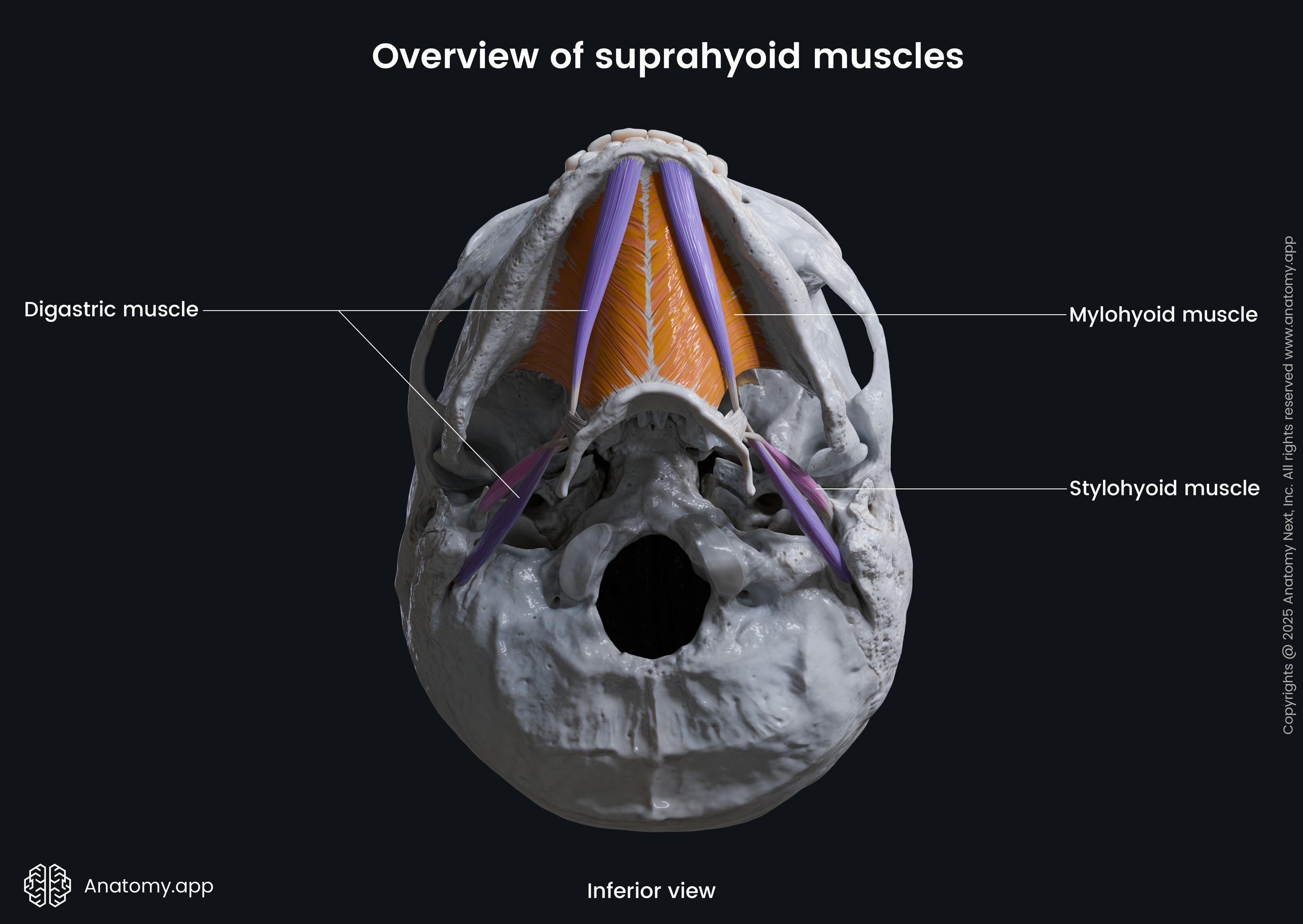
The suprahyoid group includes the following four paired muscles:
All suprahyoid muscles are named according to their origin and insertion sites, except for the digastric muscle. The first part of the name indicates the origin site, while the second part refers to the insertion of the muscle.
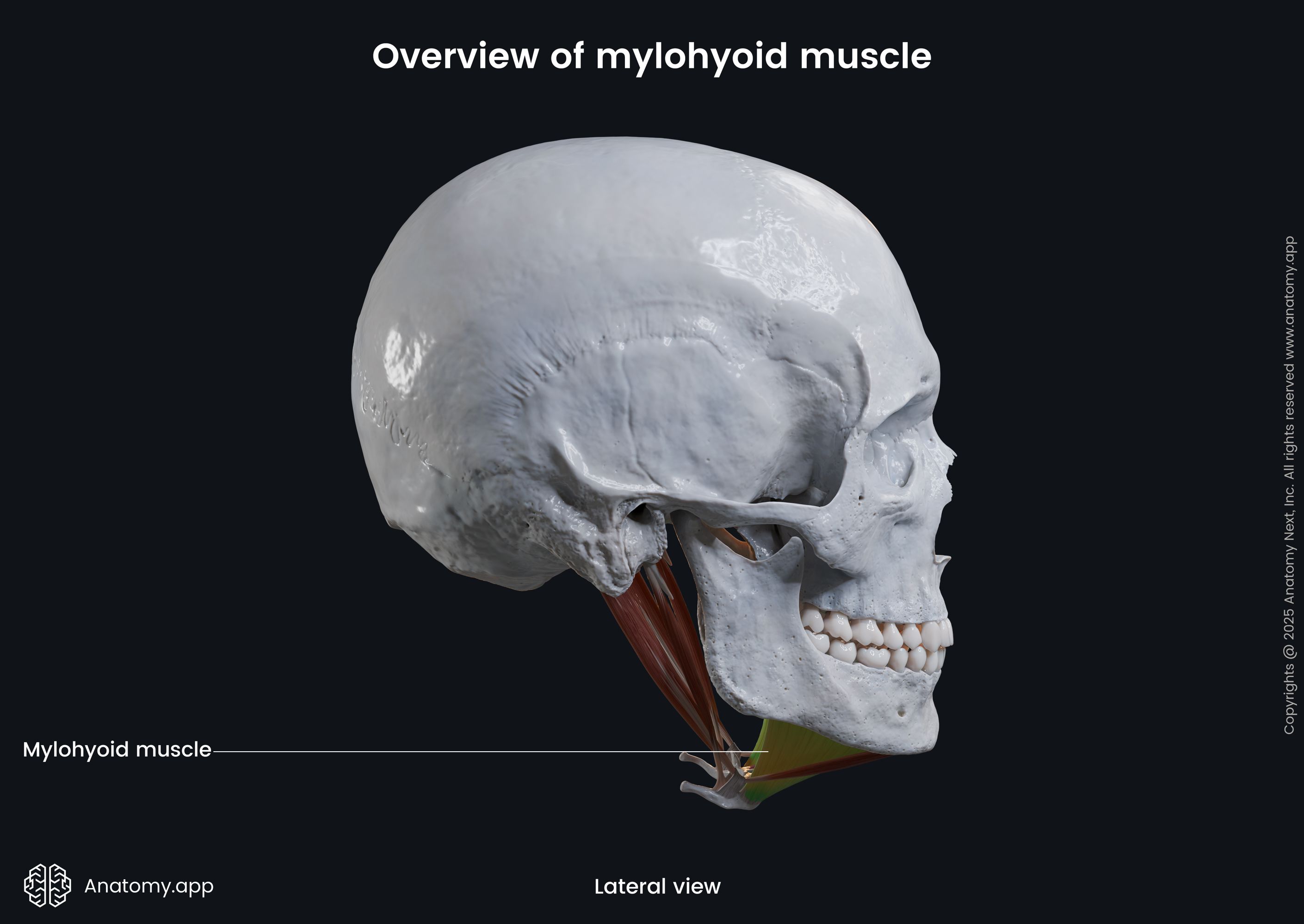
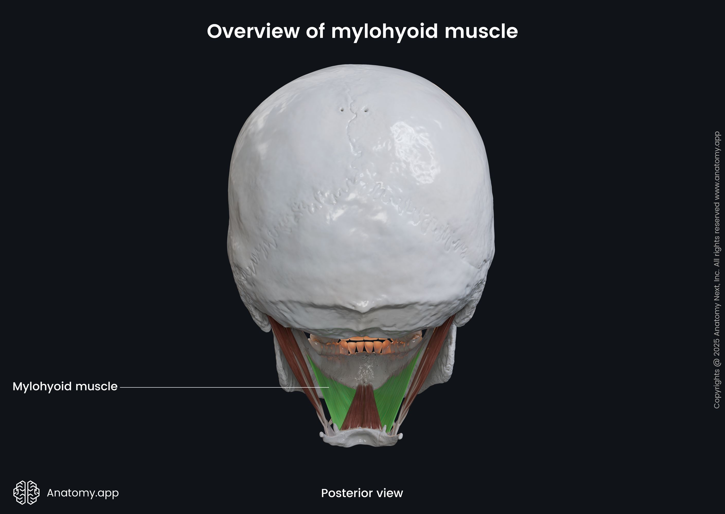
Mylohyoid
The mylohyoid is a flat, triangular muscle that forms the floor of the oral cavity and is also known as the diaphragma oris muscle. This muscle also forms the floor of the submental and submandibular triangles. It originates from the internal surface of the mandible along the mylohyoid line, stretching from the mandibular symphysis anteriorly to the last molar posteriorly. In fact, its name derives from “mylo,” the Greek word for “molar.”
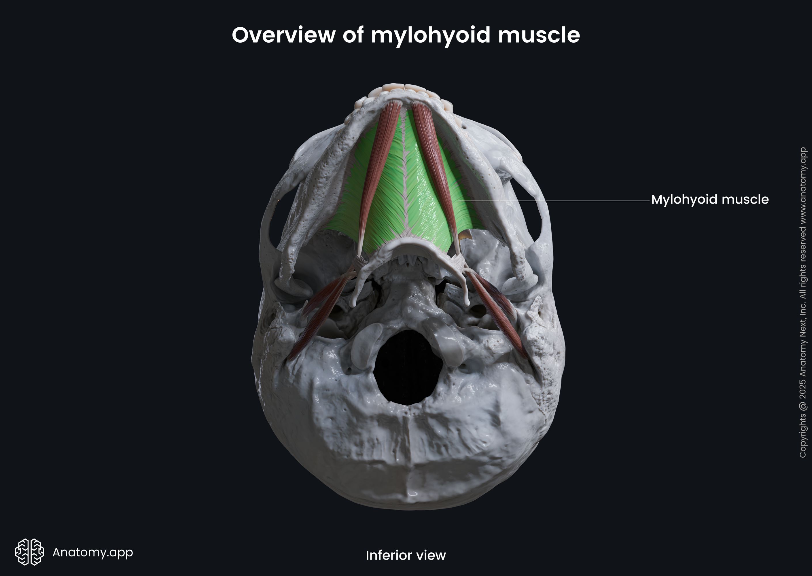
The two mylohyoid muscles meet at the midline and insert into a median fibrous raphe called the mylohyoid raphe that extends from the mandibular symphysis to the body of the hyoid bone. In addition to the mylohyoid raphe, this muscle also inserts onto the superior aspect of the body of the hyoid bone, sloping downward and medially on each side.
Geniohyoid
The geniohyoid is a relatively short, narrow muscle that is located immediately above the mylohyoid muscle. Together with the mylohyoid, it forms the floor of the oral cavity. It originates from the inferior mental spine of the mandible and inserts into the superior and anterior aspects of the body of the hyoid bone. Both geniohyoid muscles lie parallel and close to each other on either side of the midline of the floor of the oral cavity.

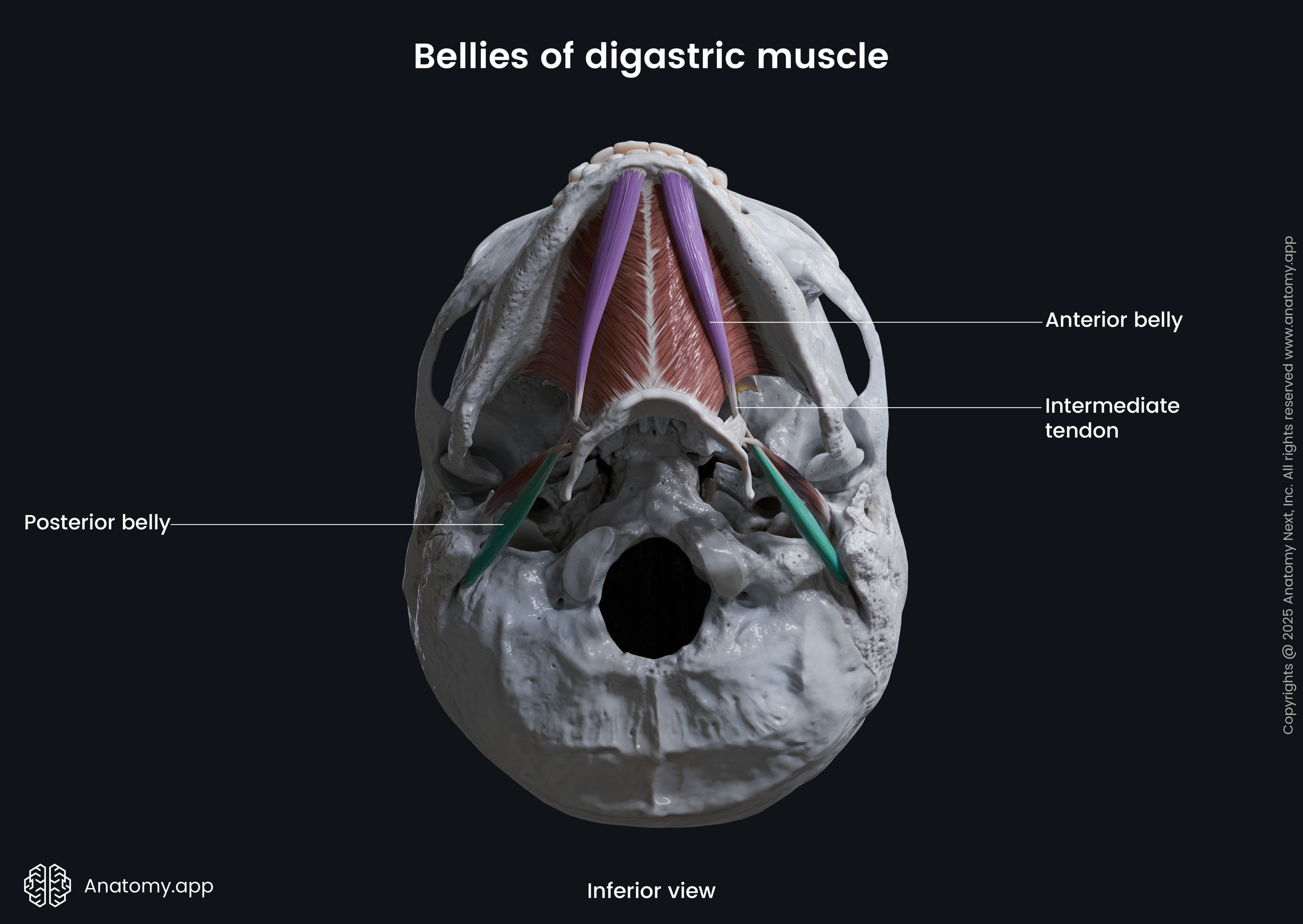
Digastric
The digastric is composed of two muscular bellies - anterior and posterior bellies. The posterior belly is relatively longer than the anterior. Both are connected by an intermediate tendon that passes through a U-shaped fibrous tissue loop that is attached to the superior aspect of the hyoid bone. The loop acts as a pulley, allowing the intermediate tendon to slide in both directions. Moreover, the intermediate tendon usually pierces the stylohyoid muscle.
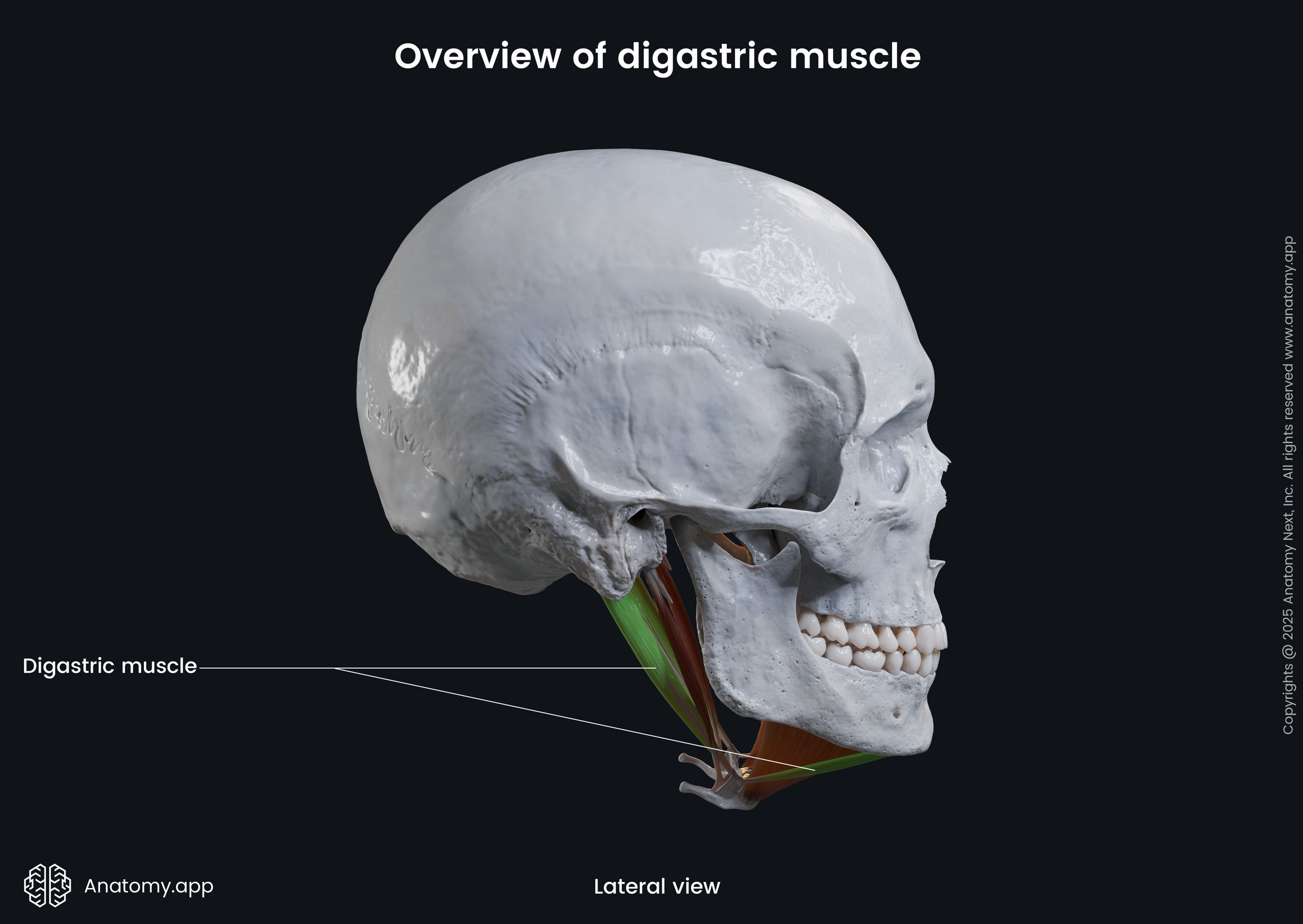
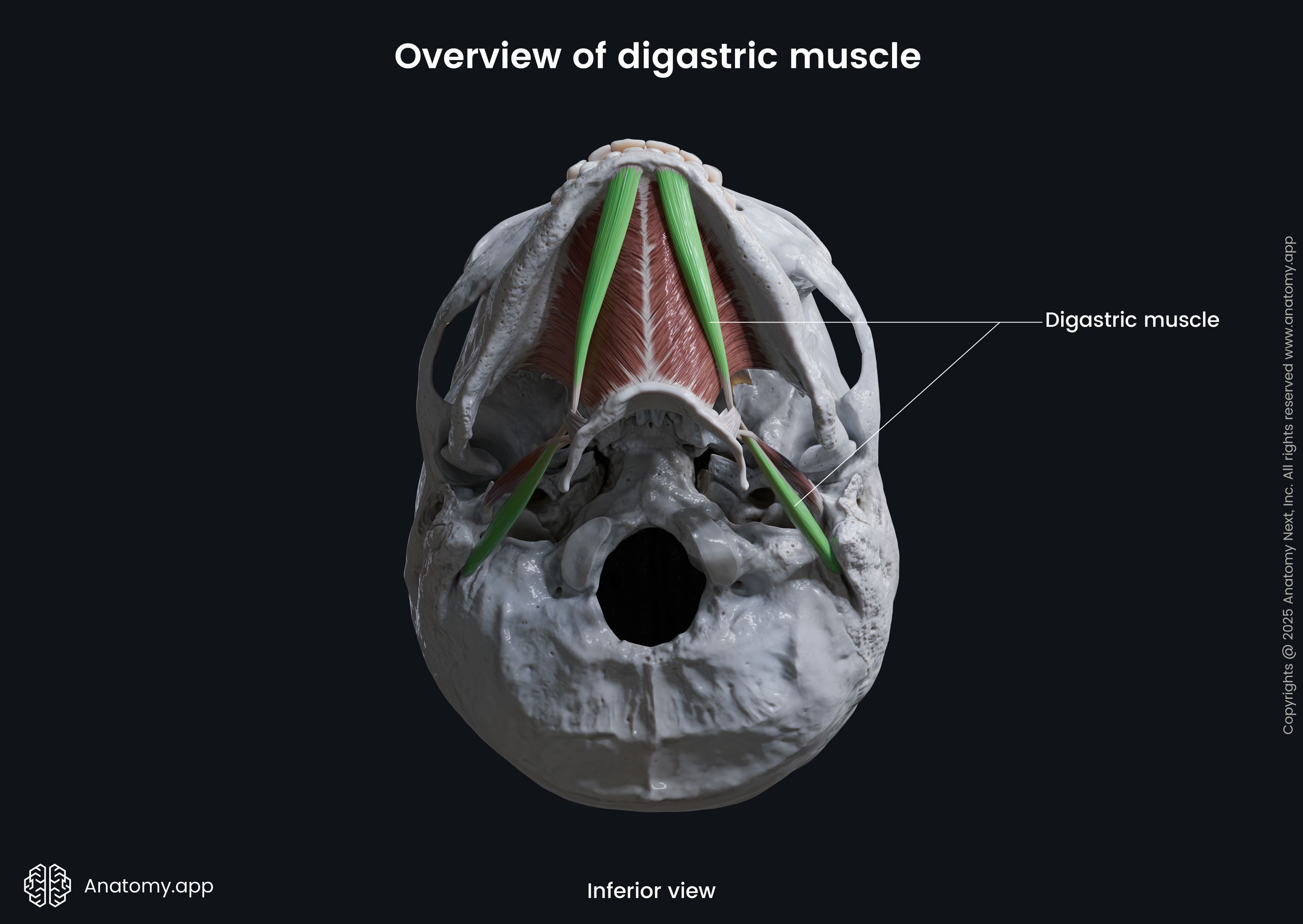
The anterior belly arises from the digastric fossa of the mandible, while the posterior belly originates from the mastoid notch of the temporal bone. Therefore, the anterior belly connects the mandible to the hyoid bone via the intermediate tendon, while the posterior belly connects the hyoid bone via the intermediate tendon to the temporal bone.
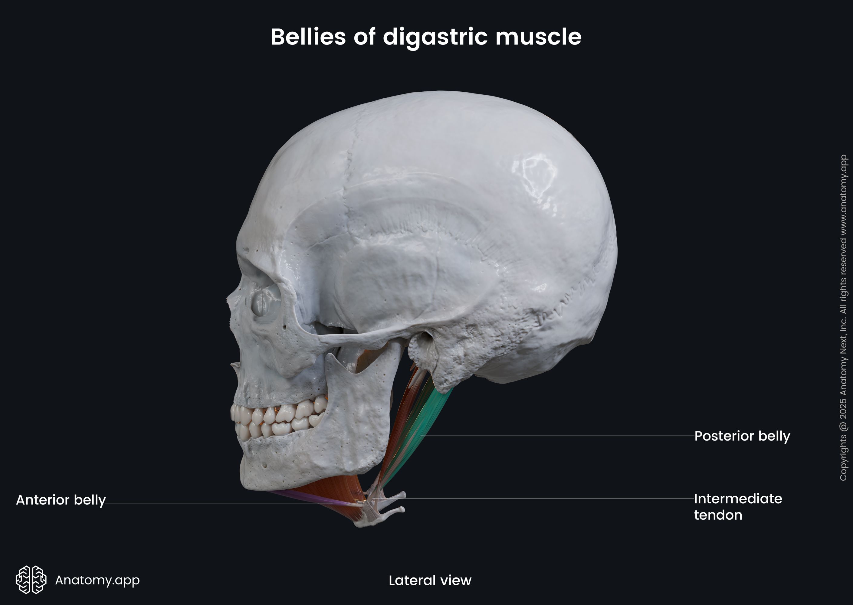
Additionally, the digastric muscle participates in forming the boundaries of three minor neck triangles that form the anterior neck triangle. The anterior belly of the muscle separates the submental and submandibular triangles. The posterior belly divides the anterior neck triangle into the submandibular triangle and the carotid triangle.
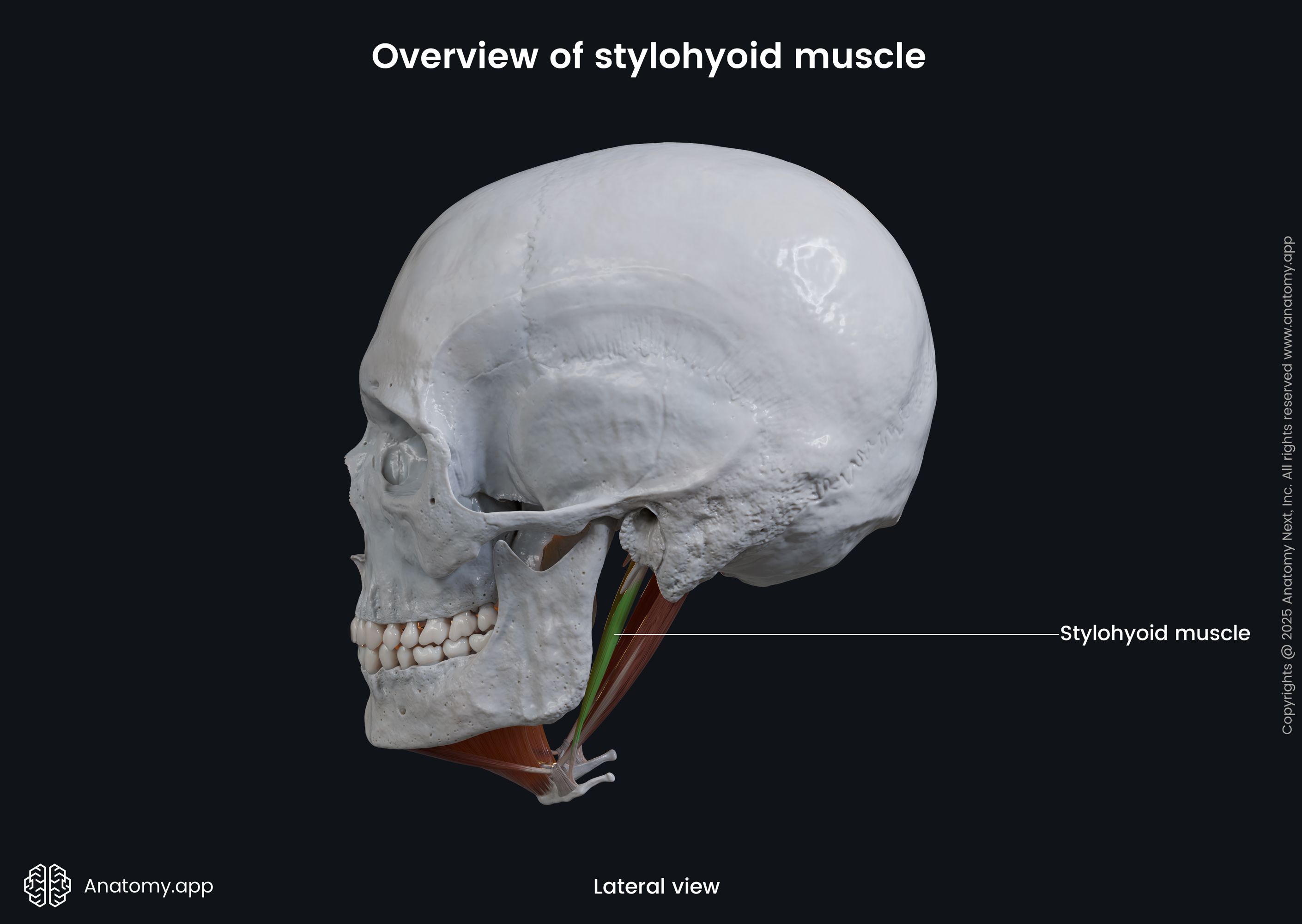
Stylohyoid
The stylohyoid is another relatively narrow muscle that is positioned posterior to the posterior belly of the digastric muscle. It arises from the styloid process of the temporal bone and inserts into the superior aspect of the hyoid bone at the junction between the greater horn and body. As mentioned before, the intermediate tendon of the digastric muscle usually penetrates the stylohyoid muscle near its insertion site.
Anatomy.app
Contact information
- For questions regarding business inquiries. Please contact:
- info@anatomy.app