- Anatomical terminology
- Skeletal system
- Joints
- Muscles
- Head muscles
- Neck muscles
- Muscles of upper limb
- Thoracic muscles
- Muscles of back
- Muscles of lower limb
- Pelvic muscles
- Muscles of thigh
- Muscles of leg
- Muscles of foot
- Heart
- Blood vessels
- Lymphatic system
- Nervous system
- Respiratory system
- Digestive system
- Urinary system
- Female reproductive system
- Male reproductive system
- Endocrine glands
- Eye
- Ear
Tensor fasciae latae
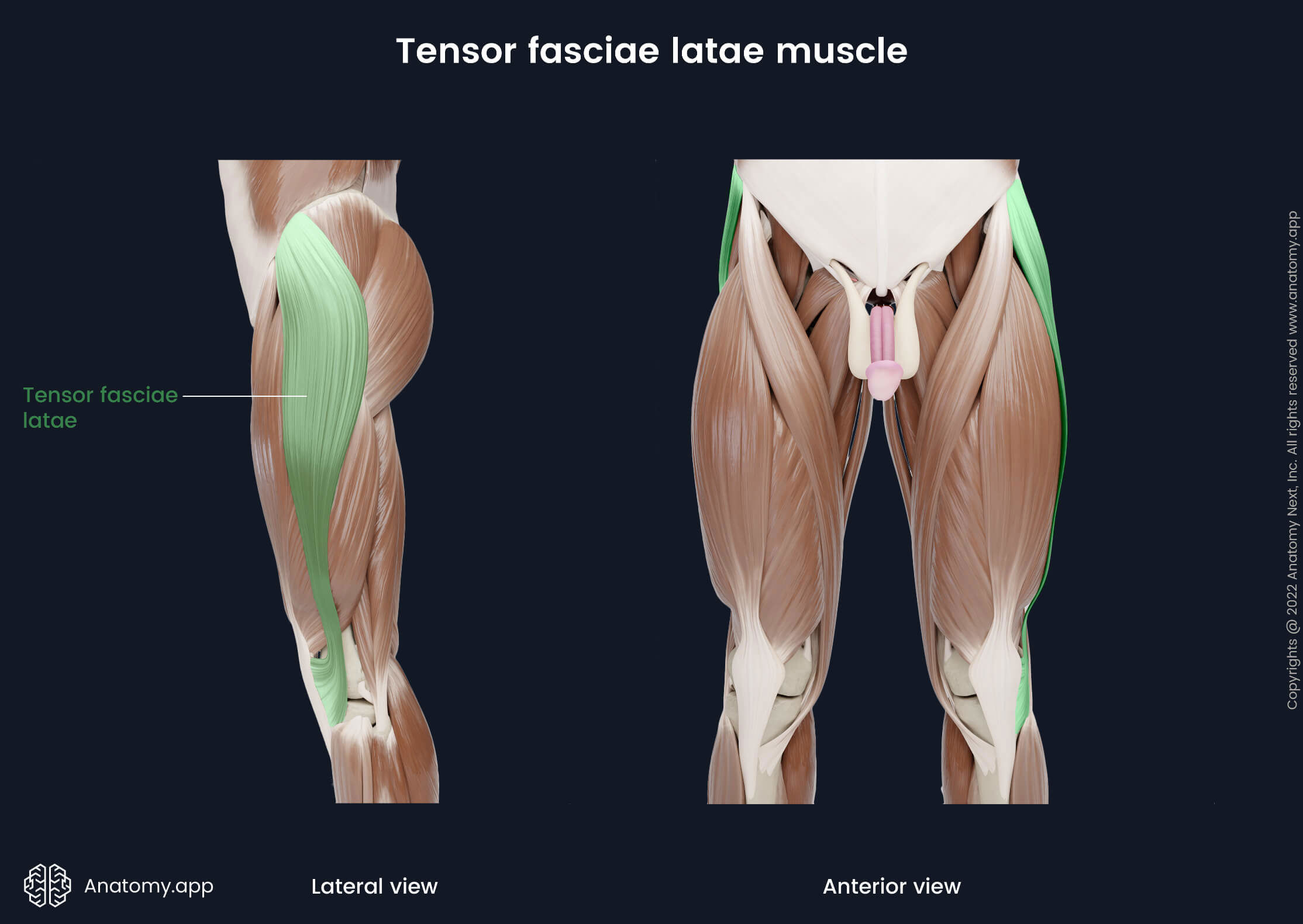
The tensor fasciae latae (Latin: musculus tensor fasciae latae) is a thin, long and superficial fusiform-shaped muscle that stretches between the ilium of the hip bone and tibia. It is situated within the gluteal region and lateral aspect of the thigh. Together with the gluteus maximus, gluteus medius and gluteus minimus muscles, the tensor fasciae latae belongs to the muscles of the gluteal region.
| Tensor fasciae latae | |
| Origin | Anterior superior iliac spine, anterior aspect of iliac crest |
| Insertion | Lateral condyle of tibia via iliotibial tract |
| Action | Internal rotation of thigh, thigh abduction, flexion of leg, leg external rotation, stabilization of hip and knee joints |
| Innervation | Superior gluteal nerve (L4 - S1) |
| Blood supply | Superior gluteal and lateral circumflex femoral arteries |

Origin
The tensor fasciae latae muscle originates from the anterior superior iliac spine and anterior aspect of the iliac crest.
Insertion
In the downward direction, the tensor fasciae latae continues as the iliotibial tract, which inserts on the lateral condyle of the tibia.
Action
The tensor fasciae latae muscle stretches the iliotibial tract and stabilizes hip and knee joints. It provides internal (medial) rotation and abduction of the thigh at the hip joint. Also, the tensor fasciae latae is responsible for flexion and external rotation of the leg at the knee joint.
Innervation
The tensor fasciae latae is innervated by the superior gluteal nerve (L4 - S1) from the sacral plexus.
Blood supply
The tensor fasciae latae muscle receives arterial blood supply from the superior gluteal and lateral circumflex femoral arteries. The first artery is a branch of the internal iliac artery, while the latter is a branch of the deep femoral artery.