- Anatomical terminology
- Skeletal system
- Joints
- Muscles
- Heart
- Blood vessels
- Blood vessels of systemic circulation
- Aorta
- Blood vessels of head and neck
- Blood vessels of upper limb
- Blood vessels of thorax
- Blood vessels of abdomen
- Arteries of abdomen
- Veins of abdomen
- Blood vessels of pelvis and lower limb
- Blood vessels of systemic circulation
- Lymphatic system
- Nervous system
- Respiratory system
- Digestive system
- Urinary system
- Female reproductive system
- Male reproductive system
- Endocrine glands
- Eye
- Ear
Portal vein
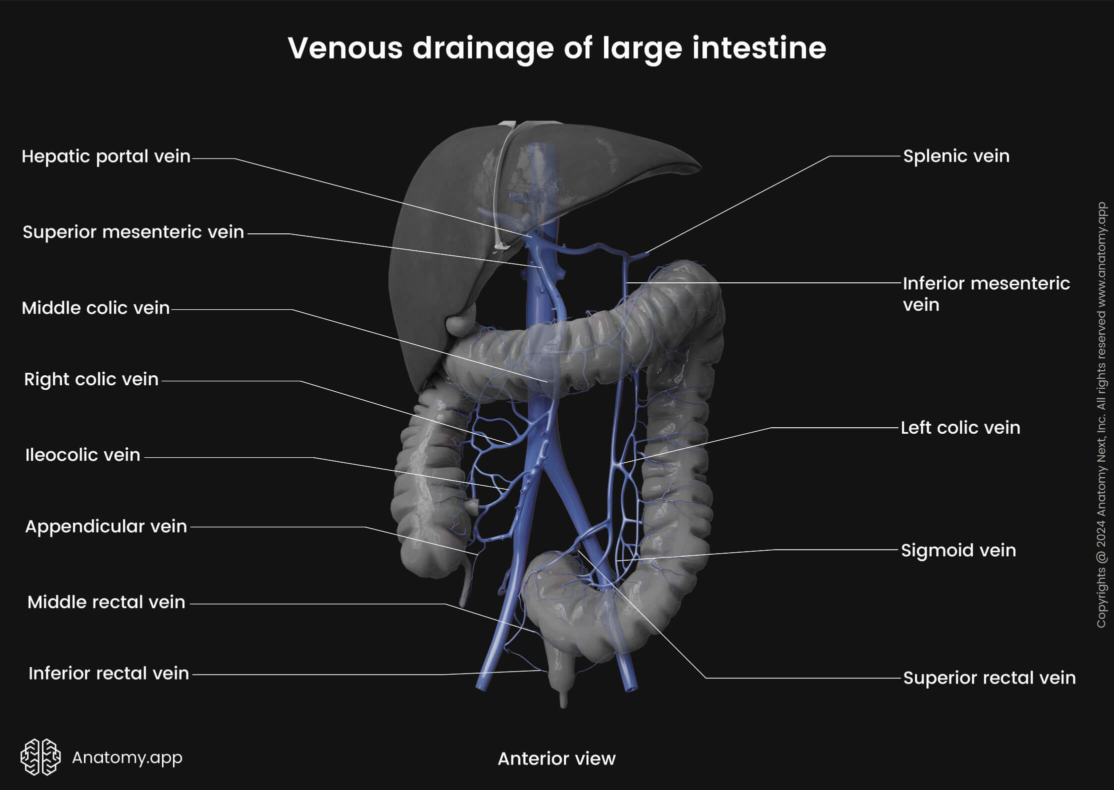
The portal vein or hepatic portal vein (Latin: vena portae hepatis) is a blood vessel located in the upper right quadrant of the abdomen that provides most of the blood supply to the liver. It is typically 8 centimeters long in adults. The portal vein is responsible for supplying the liver with nutrient-rich blood collected from the gastrointestinal tract, gallbladder, pancreas, and spleen.
The hepatic portal vein originates behind the neck of the pancreas and is usually formed by the convergence of the superior mesenteric vein and the splenic vein, referred to as the splenic-mesenteric confluence. Sometimes the hepatic portal vein also directly joins with the inferior mesenteric vein. It may also anastomose with the cystic and gastric veins.

The hepatic portal vein provides around 70% of blood supply to the liver (the hepatic arteries provide the rest). It enters the liver through the porta hepatis, which also serves as the entry point for the proper hepatic artery, and the point of exit for the bile ducts. Before reaching the liver, the portal vein divides into the right and left portal veins. Each portal vein further splits into venous branches and into portal venules, which travel along hepatic arterioles within the connective tissue between the liver lobules.
Together with a common bile duct, the portal venule and the hepatic arteriole create the hepatic portal triad. These blood vessels eventually empty into hepatic sinusoids to supply blood to the liver. After the blood is processed by the main functional liver cells (hepatocytes), it is collected by the central vein at the core of each lobule.
Blood from the central veins is ultimately collected by short right and left hepatic veins, which exit the superior surface of the liver. The hepatic veins empty into the inferior vena cava, just before the inferior vena cava penetrates the diaphragm. The inferior vena cava delivers blood to the right atrium of the heart.