- Anatomical terminology
- Skeletal system
- Joints
- Muscles
- Head muscles
-
Neck muscles
- Superficial neck muscles
- Scalene muscles
- Suprahyoid muscles
- Infrahyoid muscles
- Prevertebral muscles
- Suboccipital muscles
- Muscles of upper limb
- Thoracic muscles
- Muscles of back
- Muscles of lower limb
- Heart
- Blood vessels
- Lymphatic system
- Nervous system
- Respiratory system
- Digestive system
- Urinary system
- Female reproductive system
- Male reproductive system
- Endocrine glands
- Eye
- Ear
Rectus capitis lateralis
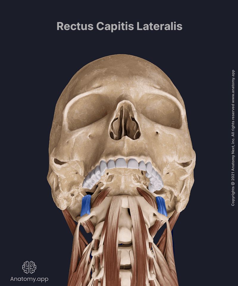
The rectus capitis lateralis (Latin: musculus rectus capitis lateralis) is a short muscle situated anterior to the spine. It extends between the atlas and the base of the skull. The rectus capitis lateralis is known as one of the prevertebral neck muscles. This muscle also belongs to the anterior neck muscles. The rectus capitis lateralis provides lateral flexion of the head.
| Rectus capitis lateralis | |
| Origin | Transverse process of atlas (C1) |
| Insertion | Jugular process of occipital bone |
| Action | Lateral flexion of head (ipsilateral), stabilization of atlanto-occipital joint |
| Innervation | Anterior rami of 1st and 2nd cervical spinal nerves (C1 - C2) |
| Blood supply | Branches of vertebral, occipital and ascending pharyngeal arteries |
Origin
The rectus capitis lateralis muscle originates from the upper surface of the transverse process of the atlas (C1).

Insertion
The rectus capitis lateralis inserts on the inferior surface of the jugular process of the occipital bone.
Action
Upon contraction, the rectus capitis lateralis muscle aids in lateral flexion of the head to the ipsilateral side. Besides the flexion, it also stabilizes the atlanto-occipital joint.
Innervation
The nerve supply to the rectus capitis lateralis is provided by the branches arising from the loop between the anterior rami of the 1st and 2nd cervical spinal nerves (C1 - C2).
Blood supply
The rectus capitis lateralis muscle receives arterial blood supply from the branches of the vertebral, occipital and ascending pharyngeal arteries. The first artery is a branch of the subclavian artery, while the occipital and ascending pharyngeal arteries are branches of the external carotid artery.