- Anatomical terminology
- Skeletal system
- Joints
- Muscles
- Head muscles
- Neck muscles
- Muscles of upper limb
- Thoracic muscles
- Muscles of back
- Muscles of lower limb
- Pelvic muscles
- Muscles of thigh
- Muscles of leg
- Muscles of foot
- Heart
- Blood vessels
- Lymphatic system
- Nervous system
- Respiratory system
- Digestive system
- Urinary system
- Female reproductive system
- Male reproductive system
- Endocrine glands
- Eye
- Ear
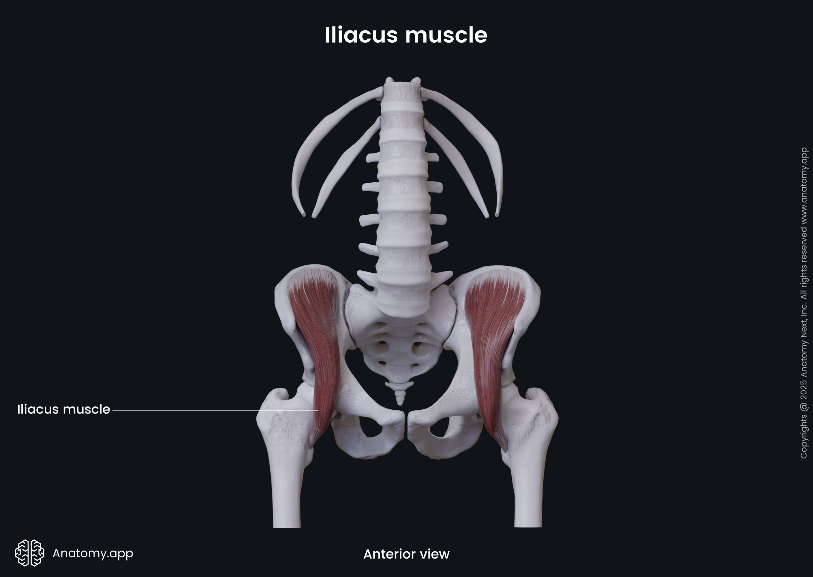
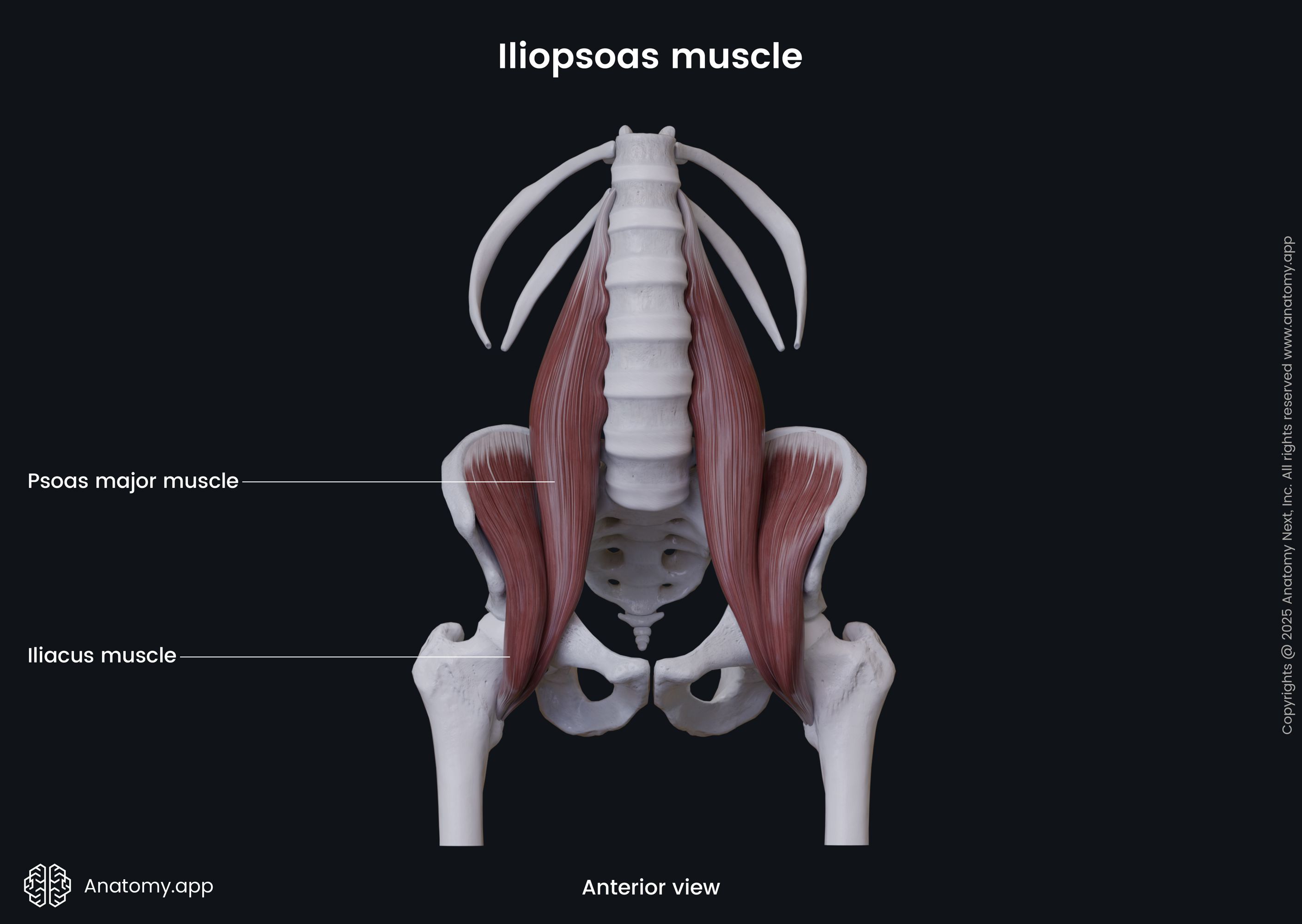
Iliacus
The iliacus (Latin: musculus iliacus) is a flat triangular-shaped muscle that is located within the pelvis and fills the iliac fossa. It stretches between the ilium of the hip bone and femur. Together with the psoas major, the iliacus forms the iliopsoas muscle. It belongs to the anterior compartment of the pelvic muscles and is one of the posterior abdominal wall muscles.
| Iliacus | |
| Origin | Iliac fossa, internal lip of iliac crest, lateral aspect of sacrum, anterior sacroiliac ligament, iliolumbar ligament |
| Insertion | Lesser trochanter of femur |
| Action | Thigh flexion, trunk flexion, thigh external rotation |
| Innervation | Femoral nerve (L2, L3) |
| Blood supply | Iliolumbar artery, branches of femoral, obturator and deep circumflex iliac arteries |
Origin
The iliacus muscle originates from the iliac fossa (upper two-thirds), internal lip of the iliac crest, lateral aspect of the sacrum, anterior sacroiliac and iliolumbar ligaments.
Insertion
The iliacus merge with the psoas major muscle and form a common tendon that inserts on the lesser trochanter of the femur.
Action
The iliacus muscle provides flexion of the thigh and trunk. Also, it assists in the external rotation of the thigh.
Innervation
The iliacus is innervated by the femoral nerve (L2, L3) that arises from the lumbar plexus.
Blood supply
The iliacus muscle is mainly supplied by the iliolumbar artery - a branch of the internal iliac artery. Besides that, it also receives arterial blood supply from the branches of the femoral, obturator and deep circumflex iliac arteries.
Anatomy.app
Contact information
- For questions regarding business inquiries. Please contact:
- info@anatomy.app