Anatomy.app Content News in October
October is here, and so is the spookiest season of the year! As the world slowly turns into a canvas of oranges, reds, and golds, typically seen only in art museums, we have prepared some ghostly updates for you. This month has been all about keeping that content A L I V E!🧟♂️
We have given a fresh look to several of our 3D models in the 3D Anatomy section. Our Media Library has also received some new videos and colorful 2D illustrations. On top of that, we have added two new 3D models to the Media Library, focusing on the nose.
So, we have rounded up everything you need to know to stay ahead in the world of content. Now, let’s dive deeper into the latest updates!
1. 3D Anatomy: Updated 3D Models
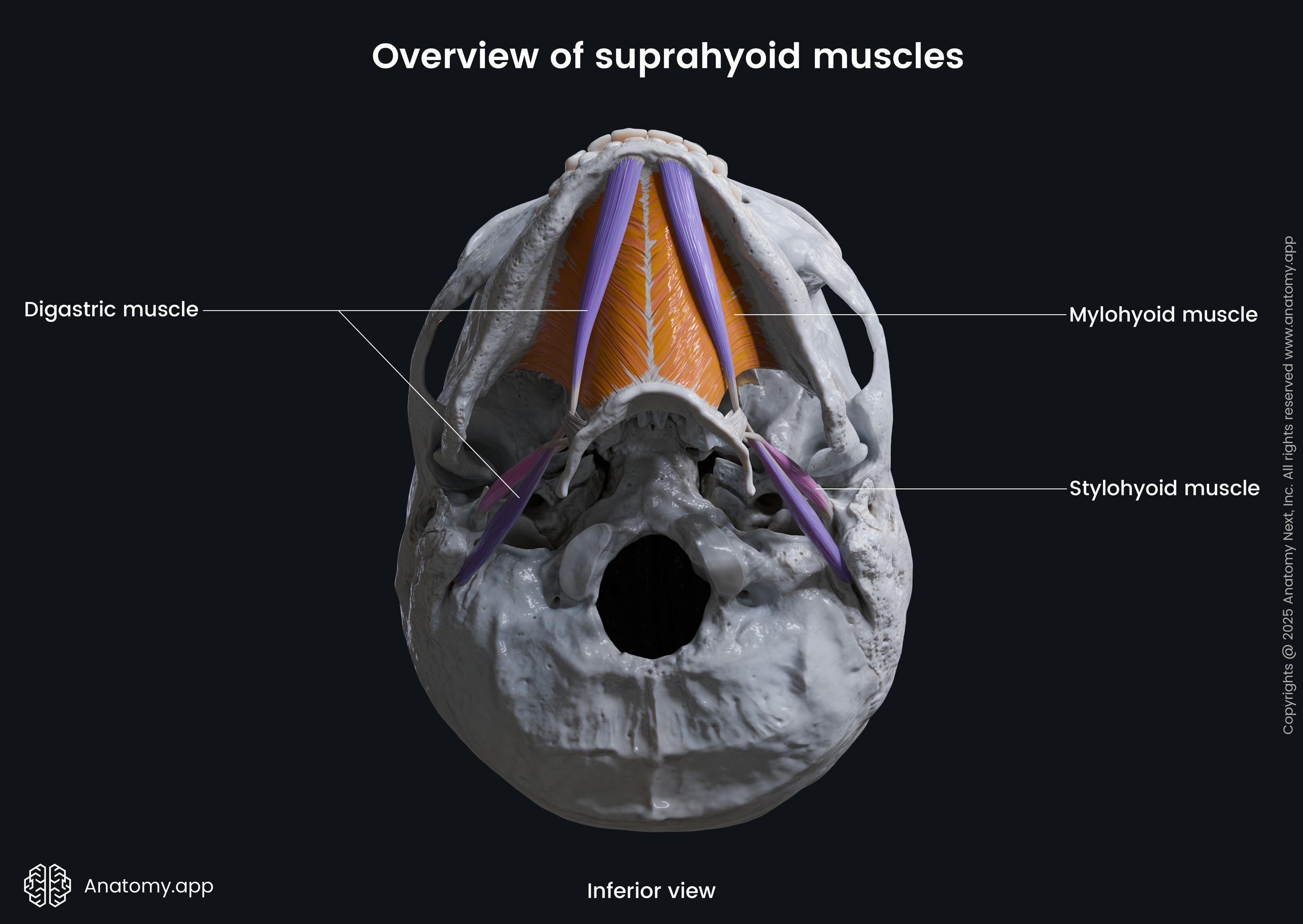
In October, we polished several of our 3D models, including all four paired muscles representing the suprahyoid neck muscles and a complete set of deep back muscles. The new back models are waiting for your inspection in the same-named article (3D Deep muscles of the back) in the 3D Spine and Back category, while you can explore the suprahyoid neck muscles in the 3D Muscles of the neck article under the 3D Head and Neck category.
Overall, we have given a makeover to 5 slides in the neck muscles article. The suprahyoid muscles are four paired muscles that are located in the anterior compartment of the neck above the hyoid bone. These muscles are attached to the superior aspect of the hyoid bone and connect it to the mandible and temporal bone. This group includes the digastric, stylohyoid, mylohyoid, and geniohyoid muscles. Together, they play an important role in chewing, swallowing, and speech.
Additionally, all 13 slides of the deep back muscles article now have a fresh look. These muscles are often overlooked and not well understood, but after reading our article and exploring our 3D models, we promise that the blurry picture will become crystal clear.
Experience the suprahyoid neck muscles in 3D: https://anatomy.app/article/muscles-of-the-neck/suprahyoid-neck-muscles
Take a closer look at the deep muscles of the back: https://anatomy.app/article/deep-muscles-of-the-back/deep-muscles-of-back-overview
2. Media Library: New 3D Models
As you may remember from one of our previous blog posts, we gave a total makeover to our 3D Facial muscles article a while ago. And while working on these 3D materials, we also revised our nasal cartilages material. Besides that, we created entirely new materials that are rarely seen or described anywhere - ta-da…the nasal ligaments!
If you want to see both models, just head to the Media Library. In the search bar, type "Nasal cartilages" or "Nasal ligaments," and you will be directed straight to the corresponding 3D models. Each model also includes a short description of the structures.
The nasal cartilages support the external framework of the nose. In our nasal cartilages model, you can explore the upper lateral cartilages, major alar cartilages, minor alar cartilages, and nasal septal cartilage.
The nasal ligaments are small but important connective structures that help maintain the shape, position, and stability of the nasal tip and sidewalls. Our model includes ligaments such as the interdomal and intercrural ligaments, pyriform ligament, longitudinal scroll ligament, and Pitanguy’s midline ligament.
Check out the nasal ligaments now: https://anatomy.app/media/nasal-ligaments-12702?categoryType=regions&searchText=nasal%20ligaments
Discover the nasal cartilages in 3D: https://anatomy.app/media/nasal-cartilages-12703?categoryType=regions&searchText=nasal%20cartilages
3. Media Library: New Videos
In addition to the updates mentioned earlier, our Media Library now includes two new videos. One is on the occipitofrontalis muscle, while the other focuses on the suprahyoid neck muscles. Both videos provide excellent insight into these muscles, making them ideal not only for students but also for clinicians or anyone looking to strengthen and deepen their knowledge of head and neck anatomy.
The video of the occipitofrontalis reviews both muscle bellies - the frontalis at the forehead and the occipitalis at the back of the head. As shown in the video, the two bellies are connected by the epicranial aponeurosis, which is a thick fibrous tissue sheath. The video provides a complete picture of how these two bellies are connected and can work together to move the scalp and eyebrows.
The video on the suprahyoid muscles is perfect for understanding their location, position, and interrelations. You can also gain insight into where each muscle is attached, as this video offers multiple angles. Overall, it gives an in-depth view of all four paired suprahyoid neck muscles.
Explore the new videos: https://anatomy.app/media?mediaType=video
4. Media Library: New Illustrations
Well, this month seems to have revolved around the suprahyoid muscles, as one more update is related to this muscle group. We have added a batch of new illustrations to our Media Library for the suprahyoid muscles. In these illustrations, you can inspect all the muscles from the lateral, posterior, and inferior views. In total, our Media Library received 21 new illustrations of these muscles.
Here is the complete list of the new illustrations added:
- Overview of the suprahyoid muscles (lateral, posterior, and inferior views)
- Geniohyoid muscle (posterior view)
- Stylohyoid muscle (lateral, posterior, and inferior views)
- Mylohyoid muscle (lateral, posterior, and inferior views)
- Digastric muscle (lateral, posterior, and inferior views)
- Bellies of the digastric muscle (lateral, posterior, and inferior views)
See the latest illustrations: https://anatomy.app/media?mediaType=image
Final Note
October indeed has its charm. And while your head is buzzing with activity, the leaves crunch underfoot, the air carries a crisp chill, cozy sweaters come out of closets, and pumpkin-spiced everything is taking over cafes. In all the study madness, don’t forget to pause for a while and enjoy it to the fullest. See you next month!
Spookily yours,
The Anatomy.app Team👻
Related blogs:
Anatomy.app
Contact information
- For questions regarding business inquiries. Please contact:
- info@anatomy.app