Gross anatomy: systemic anatomy vs regional anatomy

Some anatomical structures are extremely small and can only be observed through a microscope, while other larger structures can be seen with the naked eye. These are the two main types of anatomy – microscopic anatomy, which studies tiny anatomical structures such as tissues and cells, and gross anatomy (sometimes also called macroscopic anatomy), which studies larger structures such as bodily organs.
In this article, we will focus on gross anatomy with its different fields and explain the difference between systemic anatomy vs regional anatomy.
What is gross anatomy?
Anatomy has areas of specialization – just like most other scientific disciplines. Gross anatomy studies the larger structures of the body that are visible without the aid of magnification, for example, internal organs and external features. In Greek, “large” is macro – that is why gross anatomy is also referred to as macroscopic anatomy.
Gross anatomy is further divided into three different fields: surface or superficial anatomy, regional anatomy, and systemic anatomy.
The study of gross anatomy can be done on cadavers through dissection or with noninvasive methods through medical imaging. For example, the circulatory system can be studied by using angiography, where blood vessels are injected with an opaque dye and then visualized. Another example of noninvasive methods is radiological imaging techniques such as X-ray and MRI.
Why study gross anatomy?
Most students of health professions have to complete a dissection course in gross human anatomy. These courses aim to give you a strong understanding of basic human anatomy that can later be used to aid medical diagnosis. But since the medical school curriculum is ever-growing, gross anatomy courses sometimes don't get all the study time they deserve.
Nearly half of newly qualified doctors believe they didn't receive sufficient anatomy teaching. In response to that, some medical schools are implementing digital anatomy learning tools alongside cadaver dissections. Anatomy.app is one of them.
In fact, the combination of digital tools and gross dissection is more effective than either approach alone. Not only do they give more variety to students and professors. They also provide more flexibility to test your knowledge along the way, for example, through online quizzes. Explore the anatomy.app quizzes section!

Systemic anatomy
Systemic anatomy looks at a group of structures that work together to perform a unique body function. In other words, it focuses on whole organ systems, such as the respiratory, digestive, or nervous system.
The systemic approach allows you to focus on one type of material at a time. For example, when learning about the skeletal system, you first understand the bone structure and then move on to memorize all the bones of the human body. When you've done that, you move on to another separate system, for example, the circulatory system. This keeps the focus on one type of subject matter during the learning process.
On the downside, systemic anatomy makes it harder for you to see the connections and relationships between multiple organ systems. For example, one of the major functions of the skeletal system is to provide a rigid base for a lever system that produces movement. But the force for this comes from the muscular system. So one system is worthless without the other.
The systemic approach makes students revisit a previously learned system every time a new system is studied. When learning about the muscular system they must recall what they've learned about the skeletal system to know the muscular attachments. When learning about the nervous system, they must recall what they've learned about the muscular system so that they know which muscles are innervated by which nerves. And so on with each new system.
Digestive system anatomy – an example
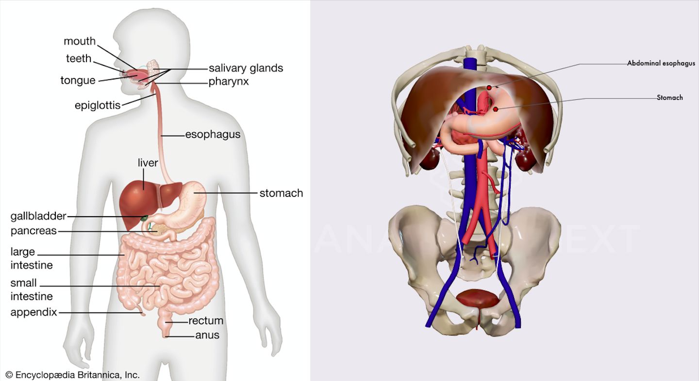
When we explore the digestive tract through the systemic approach, we concentrate on listing all the primary (mouth, pharynx, esophagus, stomach, small intestines, large intestine, rectum, and anal canal) and accessory organs (salivary glands, liver, gall bladder, pancreas) of the gastrointestinal system and describing their functions.
Moving through this system from the mouth all the way to the anus, we cover multiple regions of the human body – starting with the head and neck, moving on to the thorax and abdomen, and finishing in the pelvic region. This approach can be good to paint the big picture, but it can also feel a bit superficial. It can also be hard to visualize how the system is positioned and how it works in relation to other systems of the human body.

Regional anatomy
Regional anatomy on the other hand explains how different body structures work together in a particular region of the human body, such as the head or chest.
Regional anatomy also focuses on the diagnosis and treatment of disease or injury in the particular region that is being studied. In modern-day studies, the regional approach is used more commonly because it is easier to apply in a clinical setting than systemic anatomy.
The regional approach has many advantages – the student can focus on one region of the body at a time and thoroughly learn the muscles, nerves, vessels, etc. of the specific region. Regional anatomy also provides a better understanding of how a region functions as a unit by allowing us to explore the relationships of the various systems found there.
To compare it with the systemic approach, you are learning the bones of the region while also learning where the muscles of the region connect to these bones. At the same time, you are learning what nerves innervate the muscles of the region and what blood vessels supply the region.
The one challenge that comes with the regional approach is that it requires you to have some understanding of at least four different anatomical systems at the same time.
Gluteal region anatomy – an example
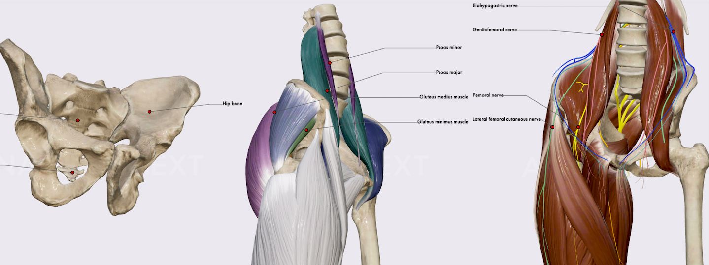
When studying the anatomy of the gluteal region, you would simultaneously learn about the bones, muscles, nerves, and vessels of the region. That is a lot to digest, so good learning materials that help you orient yourself in the wealth of information are essential.
On anatomy.app you would see that the gluteal region consists of a pair of hip bones, each of which consists of three bones - the ilium, the ischium, and the pubis.
Along with the bones, you could also explore the two groups of muscles in the hip region – anterior group or flexors (which consist of Iliopsoas (Iliacus and psoas major) and Psoas minor) and posterior group or extensors (consisting of Gluteus maximus, Gluteus medius, Gluteus minimus, Tensor fasciae latae, Piriformis, Obturator internus, Obturator externus, Superior gemellus, Inferior gemellus, and Quadratus femoris).
In the same manner, you could then explore the innervation of these muscles, along with the veins and arteries that allow blood circulation in this region.

Surface anatomy
We have looked at the two main fields of gross anatomy. Another important field is surface anatomy. It is a descriptive science that allows a surface examination of external anatomical features without dissection or radiological imaging techniques.
It is widely used to assess the position and structure of deeper organs, tissues, and systems. This can be done by studying the form and proportions of the human body as well as the anatomical landmarks which correspond to deeper structures hidden from view, both in static pose and in motion. One of the areas most frequently subjected to physical examination using auscultation or percussion is the thorax.
Foot surface anatomy – an example
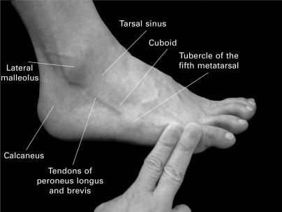
Another part of the human body where knowledge of surface anatomy is very useful is the foot. Foot issues send a lot of people to medical professionals, and since most structures in the foot are fairly superficial they can be easily palpated.
Tendons, bones, and joints tend to hurt exactly where they are injured or inflamed – that means a basic understanding of surface anatomy allows the doctor to quickly make a diagnosis or at least narrow the differential diagnosis.

In sum
Gross anatomy studies larger structures of the human body and is divided into three different fields: surface anatomy, systemic anatomy, and regional anatomy.
The best way to learn gross anatomy is through active learning techniques – and the combination of digital tools and gross dissection is more effective than either approach alone.
Systemic anatomy looks at a group of structures that work together to perform a unique body function. The systemic approach has its benefits but also makes it harder to see the connections and relationships between multiple organ systems.
Regional anatomy explains how different body structures work together in a particular region of the human body. Although it requires students to have some understanding of at least four different anatomical systems at the same time, it also provides a better understanding of how a region functions as a unit.
When comparing systemic vs regional anatomy, it is clear that each field has its usefulness, but regional anatomy is the most frequently used approach in medical schools. This approach is easier to apply in a clinical setting than systemic anatomy, especially because it makes diagnosis and treatment of disease or injury in the particular region easier.
Surface anatomy allows a surface examination of external anatomical features without invasive or radiological imaging techniques. It is especially useful for examining areas of the body where many structures are superficial, like the foot. It is also important when performing auscultation or percussion on the thorax.