Understanding Human anatomy: What is it? Types of anatomy, and how to learn anatomy better?

What is anatomy?
It's quite likely that most people have heard the word "anatomy" at least once in their lives. It could have come about by two instances - either by discussing the structure or inner workings of something or the branch of science examining the body structures of humans, animals, or other living organisms, based on dissections and the separation of body parts.
- For a precise definition, let's peek in the Merriam-Webster dictionary, which describes anatomy as:
- "a branch of morphology that deals with the structure of organisms."
- "a treatise on anatomical science and art."
- "the art of separating the parts of an organism in order to ascertain their position, relations, structure, and function (dissection)"
- "structural makeup, especially of an organism or any of its parts."
- "a separating or dividing into parts for detailed examination (f.ex the anatomy of a marriage)"
The term "anatomy" derives from the Greek word "anatomē," meaning dissection. As gathered from the definitions, it is understood as a literal dissection of a human body, animal, or other living organisms to learn about their structure or the dissection of more abstract concepts to analyze and understand them.
In this article and webpage, we are and will be explicitly concerned with human anatomy.
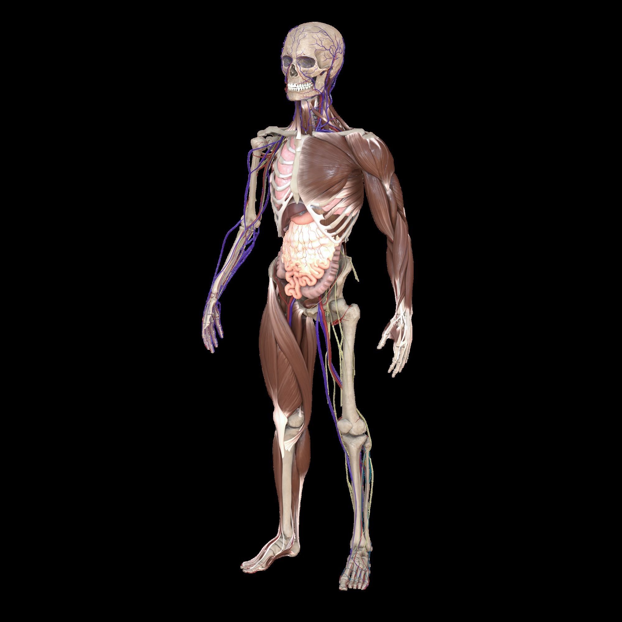
What is human anatomy?
Based on the information in the paragraphs above, we can deduce that the definition of "human anatomy" is a branch of science concerned with the human body's structural makeup and the position, relations, and functions of its parts.
Human anatomical information has historically as well as nowadays mostly been acquired through dissecting deceased humans to quite literally see how the body is made up, how specific parts are related to others, and how the body functions as a single entity.
For example, the particular muscles and their anatomy making up the upper extremity, etc.
What are the types of anatomy?
There are two types of anatomy. Macroscopic or gross anatomy and microscopic anatomy.
Macroscopic anatomy is the study of anatomical features seen by the naked eye. It includes, for example, external features or internal organs.
The second type of anatomy is microscopic anatomy. As implied by the name, this branch of anatomy is concerned with the microscopic constituents (tissues and cells) forming the larger structures. And so, microscopic anatomy relies on the usage of microscopes to study these structures.
The study of organs falls under macroscopic or gross anatomy, but the tissues and cells forming these tissues - under microscopic anatomy.
What is macroscopic or gross anatomy?
As mentioned above, macroscopic or gross anatomy is concerned with anatomical features observable by the naked eye. The main emphasis is that you don't need anything else except your eyes to gather information.
Macroscopic or gross anatomy is further subdivided into surface anatomy (the external body), regional anatomy (specific regions of the body), and systemic anatomy (specific organ systems).
Surface or superficial anatomy is concerned with the external anatomical features observable without dissection. Regional anatomy studies specific internal or external body regions and how different parts interact and cooperate in that region. Finally, systemic anatomy is the study of various organ systems and their structures—for example, the gastrointestinal or respiratory system.
What is microscopic anatomy?
Microscopic anatomy studies the microscopic constituents forming larger structures - in the level of tissues or cells. And so, microscopic anatomy relies on the usage of microscopes.
It is further subdivided into histology (tissue) and cytology (cells). Every organ is made up of tissues, and these tissues, in turn, are made up of cells.
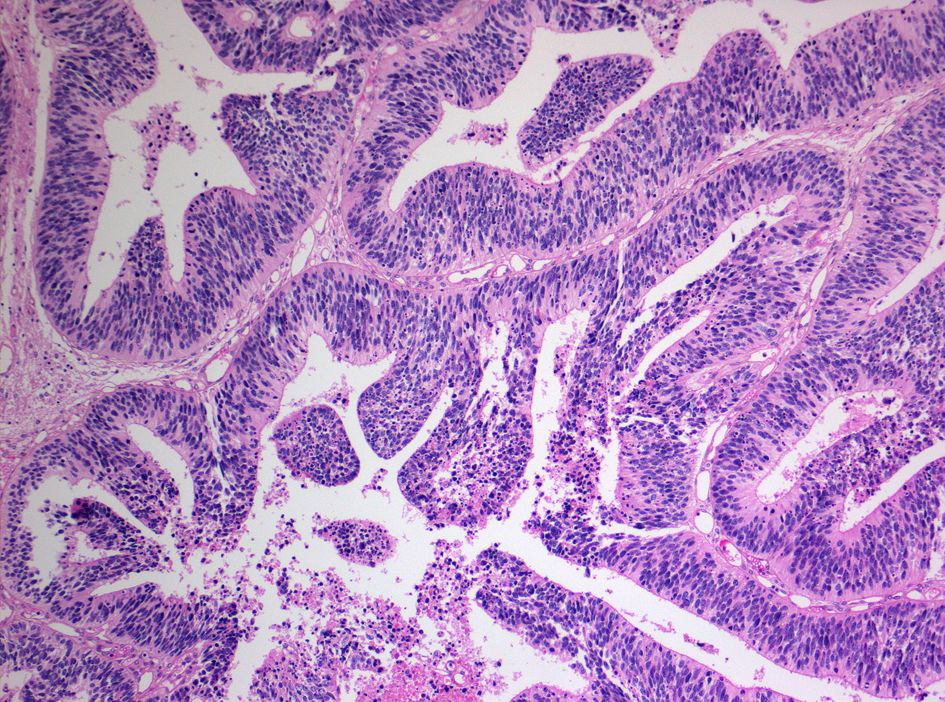
Histology is a branch of science studying the microscopic anatomy of biological tissues. It observes the correlations between structure and function. It helps in understanding certain diseases and their causes and grasp if any treatments have helped.
Specific techniques are used in order to do histological studies. Firstly, the material is fixated in chemical solutions in order to retain the natural structure. It is then cut into very thin slices using a "nano-knife."
These slices are then placed on microscope slides and colored using particular chemical agents, depending on what structures one wishes to study. One standard staining method, for example, is hematoxylin and eosin.
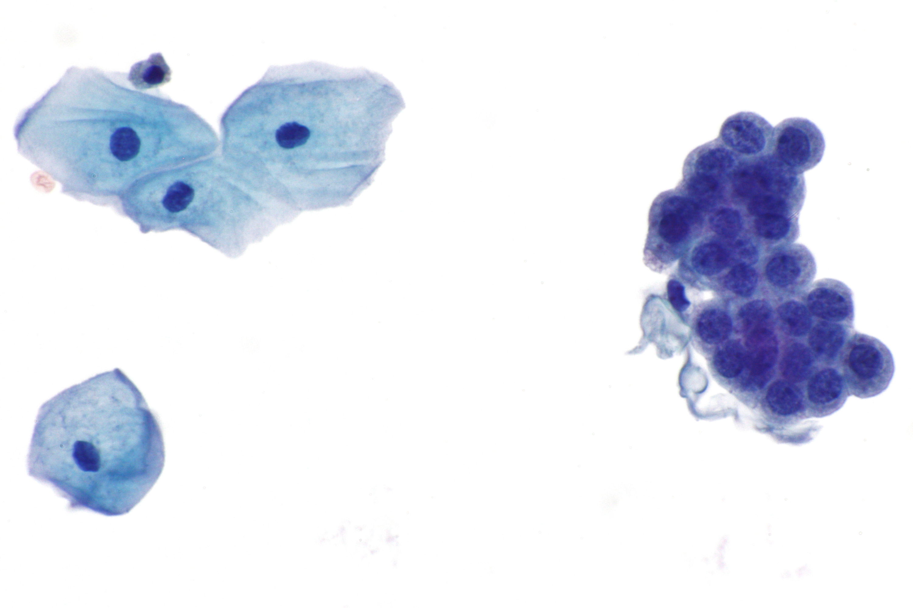
Cytology, in turn, goes even deeper, and it is concerned with examining specific cell types, often from fluid specimens. It is mostly used for cancer screening and diagnosis, especially cervical cancer (pap smear), detection of fetal abnormalities, and diagnosis of specific infectious agents.
There are two methods for obtaining samples for cytological analysis. The first, exfoliative cytology, uses cells either spontaneously shed by the body or actively scraped or brushed off from the body. It includes a pap smear, where cervical canal cells are harvested with a brush and examined for abnormalities.
The other method, intervention cytology, obtains the material by intervening in the patient's body with, for example, a needle. It includes fine-needle aspiration cytology (FNAC), where tiny amounts of the cell sample are gathered from suspicious lesions in order to rule out cancer.
What are the 5 branches of anatomy?
As seen above, anatomy divides into two broad types - macroscopic or gross anatomy and microscopic anatomy. Macroscopic anatomy is further divided into surface anatomy, regional anatomy, and systemic anatomy, but microscopic anatomy - into cytology and histology. These five subdivisions are the branches of anatomy.
But there also exist other, more specific subdivisions of anatomy. These include embryology, developmental anatomy, radiographic anatomy, and pathological anatomy.
Embryology studies the structures emerging in the period between the fertilized egg and until the 8th week in utero. It also studies gametes' (the sex cells) production and fertilization and congenital disorders occurring before birth (teratology).
Developmental anatomy studies a larger time frame because it is concerned with the development of structures starting from the fertilized egg up until the adult form.
As the term might imply, radiographic anatomy studies body structures that can be evaluated using x-rays (radiographs or CT scans).
Last but not least, pathological anatomy studies macroscopic and microscopic changes associated with diseases. It helps to observe damage caused by a specific illness and pinpoint certain causes of illness.
Why anatomy and physiology are related?
Although human anatomy is a scientific branch on its own, it most often goes hand in hand with the study of physiology. It is crucial if one wants to grasp the functioning of a human body fully. Anatomy teaches us how a body is structured, how it's held together, and how specific parts relate to others. On the other hand, physiology lets us understand what makes these parts function in the first place.
To offer an analogy, we could look at a car. The automobile's structure - its chassis, engine, brakes, axles, etc. would be the anatomy of the vehicle because it lets us understand how the car is put together and how specific parts interact.
But physiology would be the movement of the pistons in the engine and the hydraulic pressures regulating the radiator and brakes. It helps us understand why the car can move in the first place.
Knowing only the structure or only physics, we could not understand the car's whole functioning. And the same goes for the human body. To understand how we function, we need to know both human anatomy, as well as physiology.
For a complete understanding, biochemistry is also necessary. Returning to our previous analogy, this would explain why internal combustion is happening in the engine, making movement possible.
The same goes for the human body because we have particular biochemical processes occurring non-stop - formation of energy, new vital molecules, hormones, etc., necessary for a functioning body.
What are the main functions of the human body?
The human body is made up of many different “parts,” which we call organs. All these organs and their systems work together non-stop to sustain life. There is no single specific function of the human body because every part of the body contributes to this effect.
To better understand how the body does this, we need to know the specific organ systems, of which there are eleven, and their functions.
What are the 11 organ systems of the body?
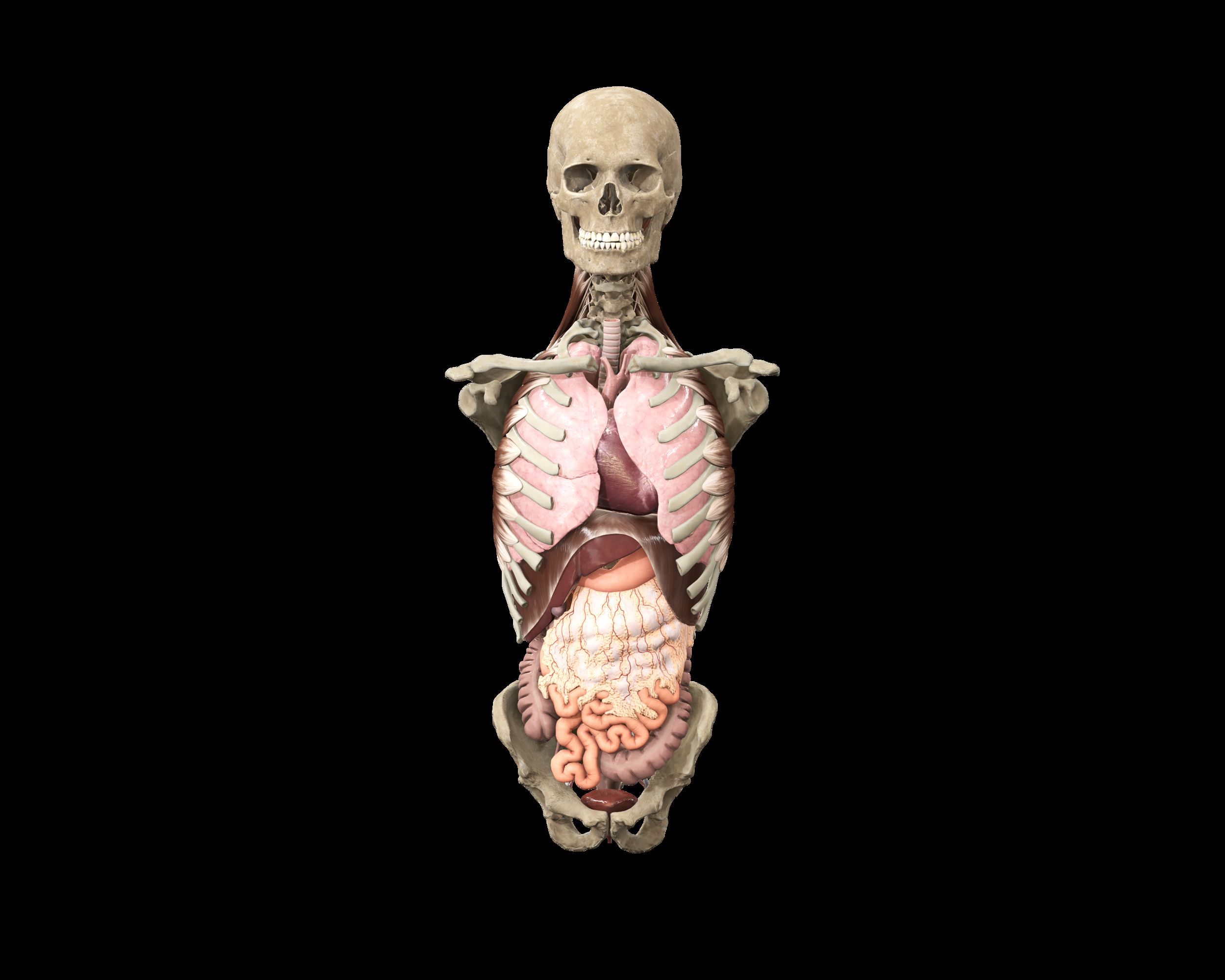
There are 11 organ systems:
- Skeletal system;
- Muscular system;
- Nervous system;
- Cardiovascular system;
- Digestive system;
- Respiratory system;
- Endocrine system;
- Renal and urinary system;
- Male and female reproductive system;
- Integumentary system;
- Immune and lymphatic system.
Each system has particular functions, and they all work together to provide reproduction and continuation of life.
The skeletal system consists of bones, tendons, ligaments, and cartilage. It forms the framework of the body, making it possible to move and stand upright. It is also involved in the storage and regulation of minerals in the blood like calcium and red blood cell production.
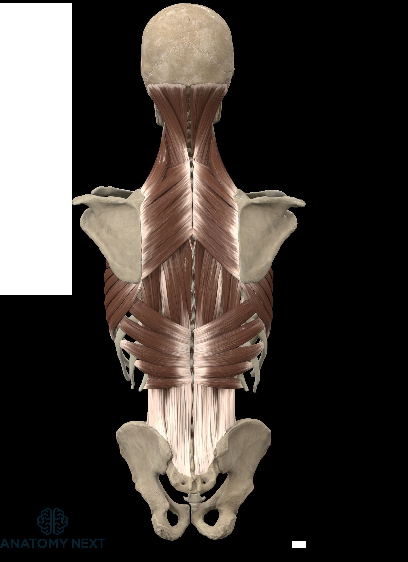
Three muscle types make up the muscular system - skeletal, smooth, and cardiac muscles. In general, the muscular system permits the body's movement, maintains proper posture, and provides blood circulation throughout the body.
The nervous system consists of the central and peripheral nervous systems. The brain and spinal cord form the central part, but all the peripheral nerves - the peripheral portion. It regulates voluntary and involuntary reactions of the human body and registers and processes sensory inputs from the environment.
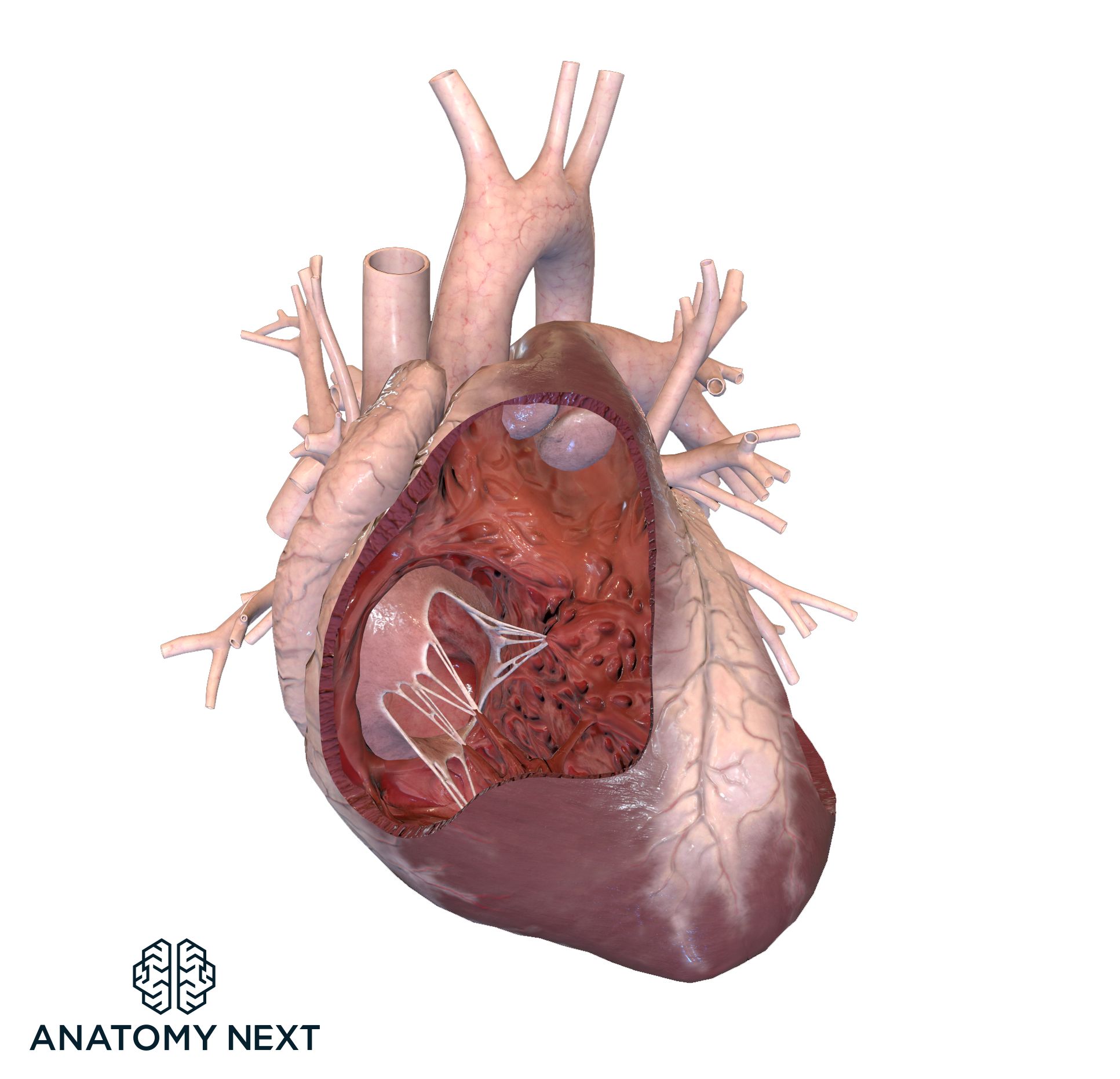
The heart, arteries, veins, and capillaries form the cardiovascular system. It provides blood circulation and transport of oxygen, carbon dioxide, nutrients, and other molecules of biological significance. It also takes part in the regulation of body temperature.
The digestive system consists of the main digestive organs - mouth, esophagus, stomach, small and large intestines, rectum, and the accessory organs - salivary glands, pancreas, liver, and gallbladder. Its primary function is the digestion and absorption of nutrients.
Formed by the nasal cavity, pharynx, larynx, trachea, bronchi, lungs, and the diaphragm, the respiratory system's primary function is gas exchange between blood and the air in the alveolar sacs.
The endocrine system consists of glands that produce specific biologically active hormones. These hormones have particular tissues of action that result in different biological adaptations.
The kidneys, ureters, bladder, and urethra make up the urinary system. This system's primary function is the filtration and excretion of excess water and biological waste products like urea. The kidneys also produce hormones that play essential roles in blood pressure regulation, the formation of red blood cells, and calcium and phosphorus concentration.
The male and female reproductive systems are very different, depending on the biological sex of the individual. Still, their main functions are the production of sex cells, fertilization, and in the case of females - nourishing and housing the fertilized egg until the fetus is ready for delivery.
The integumentary system consists of skin, hair, and nails. Its primary function is protection and regulation of body temperature, excretion of specific waste products, and sensory signals' detection through sensory receptors nested in the skin.
The lymphatic system is a part of the immune system. The lymphatic system's primary function is collecting excess fluid - or lymph, from the extracellular spaces and transporting this fluid back to the bloodstream. It also consists of lymph nodes and the spleen, which are essential members of the immune system responsible for detecting and neutralizing foreign pathogens. The primary function of the immune system is the detection and destruction of pathogens.
How many organs are there in the human body?
As we have seen, there are 11 organ systems in the human body. Different organs make up all these systems. The logical question then would be - how many individual organs are there in the body? Although this may seem straightforward, the answer is not so simple. It all depends on how one defines an organ.
For example, some specialists would count each bone as a separate organ, making up the skeletal system. But others count them as a single group - bones. It can result in huge disparities between different counting methods because only bones by themselves would add 206 separate organs.
The problem is that there is no universally accepted definition of what constitutes an organ.
One widely circulating number for the organ count is 78. This number has increased to 79 since the mesentery was added as a separate organ in 2017. This number could soon rise to 80 due to new research indicating the interstitium as a separate organ4.
But this count is not accepted unanimously by the anatomical community because there is confusion about the origin of this number.
It must be taken with a grain of salt because it falls to the eye of the beholder and how one defines an organ.
What are the 79 organs in the human body?
The number of 79 organs is very superficial, and again it must be emphasized that this count in no way is universally accepted. But due to its usage in popular culture, especially when discussing discoveries of potentially new organs, we will look at what makes up this list.
Some regions are divided in detail, such as the male and female reproductive systems, but others, like the brain, are seen as a singular organ.
It may not seem logical for some people due to the brain being a complex organ with many different parts.
As mentioned above, it would be beneficial to familiarize oneself with this count due to it being used in popular culture.
These organs are:
- Musculoskeletal system - human skeleton, joints, ligaments, skeletal muscles, and tendons.
- Digestive system - teeth, tongue, parotid, submandibular and sublingual glands, pharynx, esophagus, stomach, small intestine (duodenum, jejunum, ilium - seen as one), large intestine, rectum, liver, gallbladder, mesentery, pancreas.
- Respiratory system - nasal cavity, pharynx, larynx, trachea, bronchi, lungs, diaphragm.
- Urinary system - kidneys, ureters, bladder, urethra.
- Female reproductive system - ovaries, fallopian tubes, uterus, vagina, vulva, clitoris, placenta.
- Male reproductive system - testes, epididymis, vas deferens, seminal vesicles, prostate, bulbourethral glands, penis, scrotum.
- Endocrine system - pituitary gland, pineal gland, thyroid gland, parathyroid gland, adrenal glands, pancreas.
- Circulatory system - heart, arteries, veins, capillaries.
- Lymphatic system - lymphatic vessel, lymph node, bone marrow, thymus, spleen, tonsils.
- Nervous system - brain, medulla oblongata, pons, midbrain, cerebellum, nerves (spinal and cranial nerves - seen as one), spinal cord, ventricular system.
- Sensory organs - eyes, ears, olfactory epithelium, taste buds.
- Integumentary system - mammary glands, skin, subcutaneous tissue.
This classification will surely differ from source to source. But it is beneficial as a very rough overview of all the organs making up the human body.
Is learning anatomy hard?
The problem with anatomy is that there's just so much of it to learn. If you wish to have an in-depth understanding of how the human body works, there is no way around it. But this does not mean it is necessarily hard, although challenging.
Coming from an ex-medical student, I can say that it can be enjoyable, fun, and even easy. It's all about the routine.
Sitting down with an anatomy atlas and reading while trying to remember everything is problematic because it is a very passive process. For increased learning and retention, one needs to study actively.
It can mean underlying, writing out, looking at pictures, visualizing, or, in this case, using a specially designed program to make the process easier.
One thing is to read about a particular muscle or organ, know its theoretical location, function, etc. But it is a whole different deal to understand it visually, in 3D. Without this understanding, it can be a waste of time because anatomical knowledge without visualization in the human body's context does not give any additional practical advantage.
To become an excellent professional, one has to read and study a lot and remember and understand what has been read.
The anatomy.app provides the chance to make this process more fun, pleasant, and easy. Students can view how the learned text materializes in real-life 3D models.
Is medical school just memorization?
It would be a lie to say that there is little memorization needed to become a doctor. A large part of it is indeed pure memorization because it is necessary to remember specific terms, anatomical cues, disease names and symptoms, diagnostics, therapeutic agents and dosages, etc. But you don't have to be exceptionally gifted with memory to study medicine. It is all about finding the best routine for studying.
When it comes to anatomy, there is no denying the sheer volume of the subject. Human anatomy is very complicated. From personal experience, I can say that being able to visualize, for example, how a particular muscle is attached t.i. - it's origin and insertion can make it way easier to remember it later and remember the function of that muscle.
Suppose you can visualize the material as you are learning it in real-time. In that case, your understanding and comprehension will increase, making it easier to understand and remember it in the long term. That is why anatomy.app gives its users an advantage.
How can I memorize anatomy quickly?
You can't. The reality is that speed decreases comprehension and retention. There are no quick fixes to memorize anatomy. Or if you do learn it in a very short period for an exam, let's say, you will forget it very soon after. It is not ideal for future medical professionals.
But there are specific methods of memorizing anatomical information easier. For example, using a visual aid program like anatomy.app, because visual learning is active learning, increasing retention. Ideally, you should combine the usage of anatomy.app with specific memory techniques, like the "memory palace" or "loci method," to remember it better. We will return to these techniques in a subsequent blog post.
